File:IPLab8HBV9.jpg
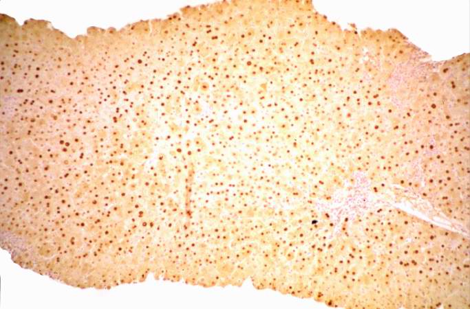
Revision as of 02:50, 21 August 2013 by Seung Park (talk | contribs) (This is a low-power photomicrograph of the same liver reacted with antibody specific for hepatitis B core antigen (HBcAg). The hepatocytes that contain HBcAg stain brown. Note that even at this low magnification, many brown-staining nuclei can be seen.)
No higher resolution available.
IPLab8HBV9.jpg (683 × 450 pixels, file size: 51 KB, MIME type: image/jpeg)
This is a low-power photomicrograph of the same liver reacted with antibody specific for hepatitis B core antigen (HBcAg). The hepatocytes that contain HBcAg stain brown. Note that even at this low magnification, many brown-staining nuclei can be seen.
File history
Click on a date/time to view the file as it appeared at that time.
| Date/Time | Thumbnail | Dimensions | User | Comment | |
|---|---|---|---|---|---|
| current | 02:50, 21 August 2013 | | 683 × 450 (51 KB) | Seung Park (talk | contribs) | This is a low-power photomicrograph of the same liver reacted with antibody specific for hepatitis B core antigen (HBcAg). The hepatocytes that contain HBcAg stain brown. Note that even at this low magnification, many brown-staining nuclei can be seen. |
- You cannot overwrite this file.
File usage
The following page links to this file: