File:IPLab8HBV1.jpg
Revision as of 02:47, 21 August 2013 by Seung Park (talk | contribs) (This is a low-power photomicrograph of liver from this case. This section was stained with a modified aldehyde fuchsin and counterstained with hematoxylin and eosin. Modified aldehyde fuchsin colors cystine-rich proteins--such as HBsAg and elastic fibe...)
No higher resolution available.
IPLab8HBV1.jpg (685 × 450 pixels, file size: 22 KB, MIME type: image/jpeg)
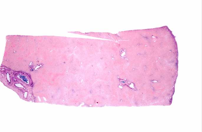
This is a low-power photomicrograph of liver from this case. This section was stained with a modified aldehyde fuchsin and counterstained with hematoxylin and eosin. Modified aldehyde fuchsin colors cystine-rich proteins--such as HBsAg and elastic fibers--deep purple. The cytoplasm of most liver cells (and RBCs) stain red due to the eosin and have dark blue nuclei.
File history
Click on a date/time to view the file as it appeared at that time.
| Date/Time | Thumbnail | Dimensions | User | Comment | |
|---|---|---|---|---|---|
| current | 02:47, 21 August 2013 | | 685 × 450 (22 KB) | Seung Park (talk | contribs) | This is a low-power photomicrograph of liver from this case. This section was stained with a modified aldehyde fuchsin and counterstained with hematoxylin and eosin. Modified aldehyde fuchsin colors cystine-rich proteins--such as HBsAg and elastic fibe... |
- You cannot overwrite this file.
File usage
The following page links to this file: