File:IPLab7Osteosarcoma6.jpg
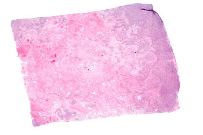
Revision as of 02:14, 21 August 2013 by Seung Park (talk | contribs) (This is a low-power photomicrograph of decalcified histologic section from this tumor. Note the blue color (cell nuclei stain blue) of much of this section indicating the increased cellularity of the tumor.)
No higher resolution available.
IPLab7Osteosarcoma6.jpg (675 × 450 pixels, file size: 28 KB, MIME type: image/jpeg)
This is a low-power photomicrograph of decalcified histologic section from this tumor. Note the blue color (cell nuclei stain blue) of much of this section indicating the increased cellularity of the tumor.
File history
Click on a date/time to view the file as it appeared at that time.
| Date/Time | Thumbnail | Dimensions | User | Comment | |
|---|---|---|---|---|---|
| current | 02:14, 21 August 2013 | | 675 × 450 (28 KB) | Seung Park (talk | contribs) | This is a low-power photomicrograph of decalcified histologic section from this tumor. Note the blue color (cell nuclei stain blue) of much of this section indicating the increased cellularity of the tumor. |
- You cannot overwrite this file.
File usage
The following page links to this file: