File:IPLab7Osteosarcoma5.jpg
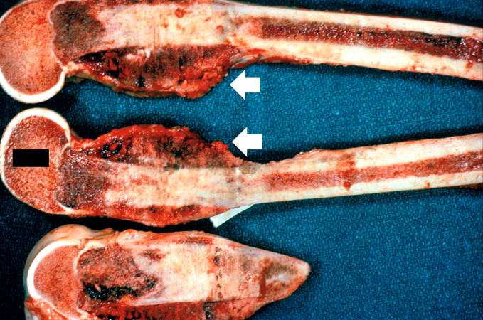
Revision as of 02:14, 21 August 2013 by Seung Park (talk | contribs) (These are cut sections of the distal femur containing the tumor. The periosteal involvement is evident from this picture (arrows).)
No higher resolution available.
IPLab7Osteosarcoma5.jpg (679 × 450 pixels, file size: 67 KB, MIME type: image/jpeg)
These are cut sections of the distal femur containing the tumor. The periosteal involvement is evident from this picture (arrows).
File history
Click on a date/time to view the file as it appeared at that time.
| Date/Time | Thumbnail | Dimensions | User | Comment | |
|---|---|---|---|---|---|
| current | 02:14, 21 August 2013 | | 679 × 450 (67 KB) | Seung Park (talk | contribs) | These are cut sections of the distal femur containing the tumor. The periosteal involvement is evident from this picture (arrows). |
- You cannot overwrite this file.
File usage
The following page links to this file: