File:IPLab6SenileAmyloidosis1.jpg
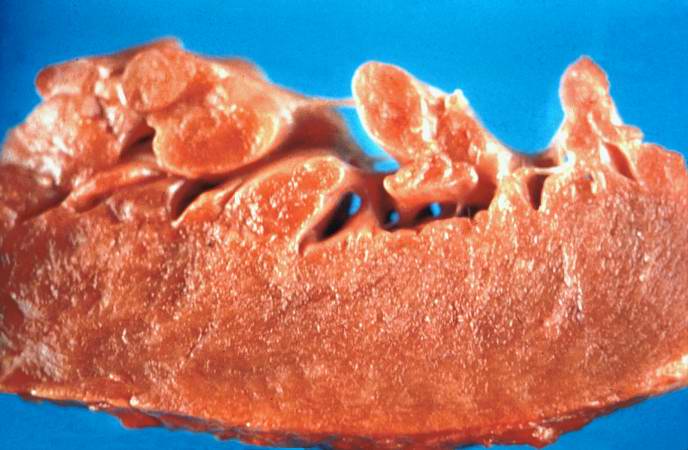
Revision as of 20:24, 20 August 2013 by Seung Park (talk | contribs) (This is a gross photograph of section of heart tissue from this case. The tissue was firm and had a waxy texture. If you use your imagination you can see pale yellow areas within this tissue which represent the amyloid deposits.)
No higher resolution available.
IPLab6SenileAmyloidosis1.jpg (688 × 450 pixels, file size: 47 KB, MIME type: image/jpeg)
This is a gross photograph of section of heart tissue from this case. The tissue was firm and had a waxy texture. If you use your imagination you can see pale yellow areas within this tissue which represent the amyloid deposits.
File history
Click on a date/time to view the file as it appeared at that time.
| Date/Time | Thumbnail | Dimensions | User | Comment | |
|---|---|---|---|---|---|
| current | 20:24, 20 August 2013 | | 688 × 450 (47 KB) | Seung Park (talk | contribs) | This is a gross photograph of section of heart tissue from this case. The tissue was firm and had a waxy texture. If you use your imagination you can see pale yellow areas within this tissue which represent the amyloid deposits. |
- You cannot overwrite this file.
File usage
The following page links to this file: