File:IPLab5Downs2.jpg
Revision as of 18:27, 19 August 2013 by Seung Park (talk | contribs) (These cells, obtained by amniocentesis, were cultured and then arrested in metaphase. Nuclei from these cells were isolated and stained to demonstrate the banding pattern of each chromosome. This photograph shows a "chromosome spread." Each chromosome ...)
No higher resolution available.
IPLab5Downs2.jpg (599 × 450 pixels, file size: 17 KB, MIME type: image/jpeg)
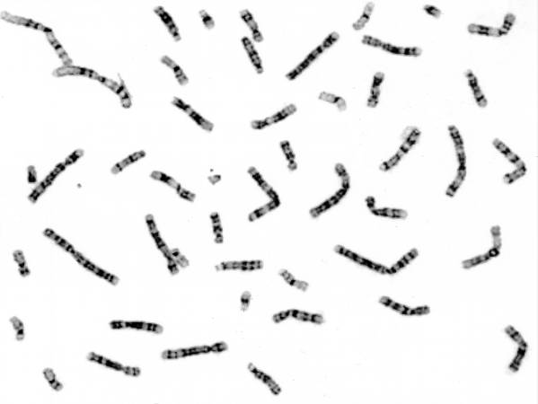
These cells, obtained by amniocentesis, were cultured and then arrested in metaphase. Nuclei from these cells were isolated and stained to demonstrate the banding pattern of each chromosome. This photograph shows a "chromosome spread." Each chromosome is identified and lined up to give a karyotype (next page).
Amniocentesis is a procedure in which a needle is inserted transabdominally through the uterus, into the amniotic sac, and amniotic fluid is withdrawn.
File history
Click on a date/time to view the file as it appeared at that time.
| Date/Time | Thumbnail | Dimensions | User | Comment | |
|---|---|---|---|---|---|
| current | 18:27, 19 August 2013 | | 599 × 450 (17 KB) | Seung Park (talk | contribs) | These cells, obtained by amniocentesis, were cultured and then arrested in metaphase. Nuclei from these cells were isolated and stained to demonstrate the banding pattern of each chromosome. This photograph shows a "chromosome spread." Each chromosome ... |
- You cannot overwrite this file.
File usage
The following page links to this file: