File:IPLab2FattyChange11.jpg
No higher resolution available.
IPLab2FattyChange11.jpg (675 × 450 pixels, file size: 58 KB, MIME type: image/jpeg)
File history
Click on a date/time to view the file as it appeared at that time.
| Date/Time | Thumbnail | Dimensions | User | Comment | |
|---|---|---|---|---|---|
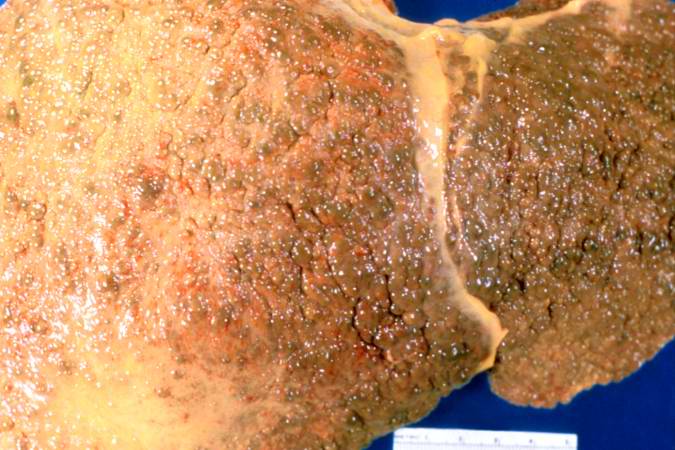
| current | 12:00, 19 August 2013 | | 675 × 450 (58 KB) | Peter Anderson (talk | contribs) | This gross photograph of liver demonstrates severe nodular cirrhosis. Note the extensive scarring of the capsule and the nodular projections of tissue through the uncut capsule in this tissue. The green color is due to the accumulation of bile pigment. |
You cannot overwrite this file.
File usage
The following page uses this file:
