IPLab:Lab 4:Septic Emboli
Images
-
 This gross photograph of lung demonstrates microabscesses due to septic embolization. Note the small 2 to 3-mm lesions scattered throughout this lung tissue (arrows).
 -
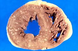 This is a gross photograph of myocardium with multiple embolic lesions scattered throughout the left and right ventricles.
 -
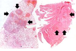 This low-power photomicrograph shows a section of lung on the left and myocardium on the right. Both pieces of tissue have multiple embolic lesions seen as blue staining areas with massive infiltration of inflammatory cells (arrows).
 -
-
-

-
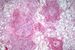
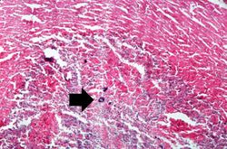 This is a higher-power photomicrograph of the abscess within the myocardium illustrating colonies of bacteria as the dark blue-staining material (arrow).
 -
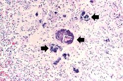
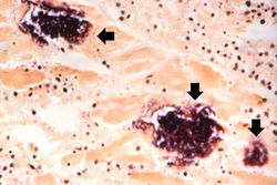 This is a high-powered photomicrograph of a myocardial abscess stained with a special tissue Gram stain (Brown & Brenn) to illustrate the colonies of bacteria in this myocardial tissue (arrows).


