File:IPLab2Atrophy9.jpg
No higher resolution available.
IPLab2Atrophy9.jpg (670 × 450 pixels, file size: 47 KB, MIME type: image/jpeg)
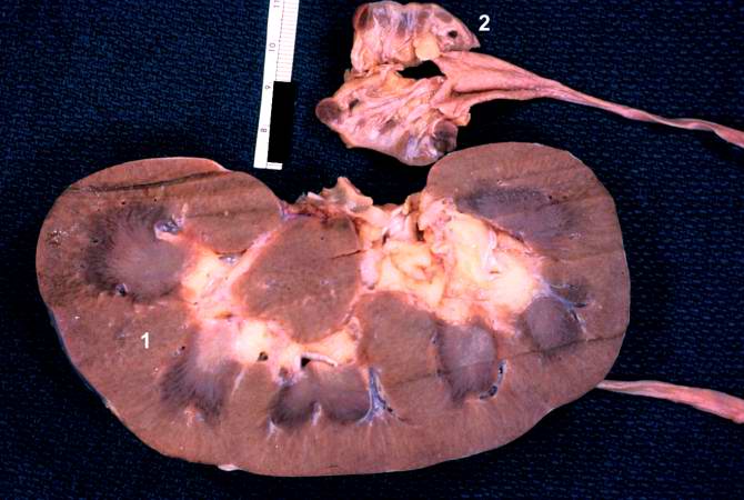
The two kidneys in this slide are from the same patient. One kidney (1) is relatively normal, although increased in size due to compensatory hypertrophy. The other kidney (2) is very small with only rudimentary nodules of renal parenchyma. This kidney had never developed and therefore this process represents hypoplasia. How does one differentiate between atrophy and hypoplasia?
File history
Click on a date/time to view the file as it appeared at that time.
| Date/Time | Thumbnail | Dimensions | User | Comment | |
|---|---|---|---|---|---|
| current | 11:11, 19 August 2013 | | 670 × 450 (47 KB) | Peter Anderson (talk | contribs) | The two kidneys in this slide are from the same patient. One kidney (1) is relatively normal, although increased in size due to compensatory hypertrophy. The other kidney (2) is very small with only rudimentary nodules of renal parenchyma. This kidney ... |
You cannot overwrite this file.
File usage
The following page uses this file:
