IPLab:Lab 13:Myelomeningocele
Clinical Summary
This male fetus of a 21-year-old Rh-positive mother (Gravida 2 Para 1011) was the product of an induced delivery at 21 weeks gestation. At 19 weeks gestation, routine prenatal examination had revealed a maternal serum alpha-fetoprotein (AFP) level that was 4.2 times the mean for a mother at this point in gestation. A targeted ultrasound was ordered--which revealed a lumbosacral neural tube defect--and an amniocentesis was performed. Amniotic fluid showed an AFP level that was 5.3 times the calculated median value and cytogenetic analysis demonstrated a normal male (46XY) chromosome pattern. The parents requested termination of the pregnancy and a 465-gram male nondysmorphic stillborn fetus was delivered vaginally.
At autopsy there was a myelomeningocele in the lumbar region that measured 1.6 cm in width and 3.7 cm in length.
Images
-
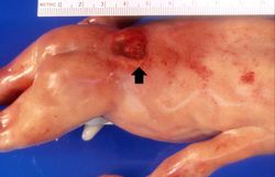 This is a gross photograph of the fetus at autopsy. Note the defect in the lower lumbar region of the spinal column (arrow). The myelomeningocele can be seen protruding from this defect.
 -
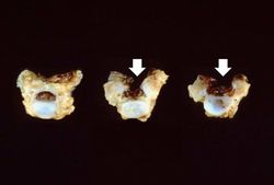 This gross photograph shows consecutive lumbar vertebra from this case. Note the defect (arrows) in the two vertebral bodies on the right. This defect was caused by failure of the vertebral column to properly close.
 -
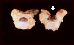 This is a closer view of the previous gross photograph showing a normal lumbar vertebra from this case on the left. Once again, note the defect (arrow) in the vertebral body on the right due to failure of the vertebral column to close properly.
 -
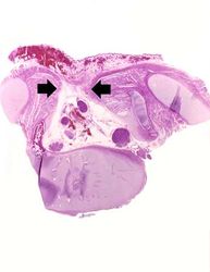 This is a low-power photomicrograph of one of the vertebral bodies from this case. In this section there are defects (arrows) in the vertebral body but the skin can be seen over the open vertebral canal.
 -
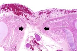 This is a higher-power photomicrograph of one of the vertebral bodies from this case. The defect (arrows) in the vertebral body is seen more clearly. The spinal cord is disrupted and there are areas of hemorrhage in this region.
 -
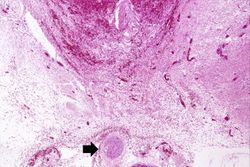 This is a high-power photomicrograph of the spinal cord (arrow) immediately beneath the area of hemorrhage.
 -
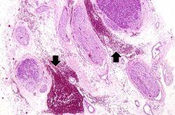 This high-power photomicrograph of the spinal cord within the vertebral column shows the hemorrhage (arrows) in this region.


Study Questions
Additional Resources
Reference
- eMedicine Medical Library: Spina Bifida
- eMedicine Medical Library: Neural Tube Defects in the Neonatal Period
- eMedicine Medical Library: Neural Tube Defects
- Merck Manual: Multifactorial (Complex) Inheritance
- Merck Manual: Spina Bifida
Journal Articles
- Cortes RA, Farmer DL. Recent advances in fetal surgery. Semin Perinatol 2004 Jun;28(3):199-211.