File:IPLab3Bronchopneumonia7.jpg
IPLab3Bronchopneumonia7.jpg (668 × 450 pixels, file size: 86 KB, MIME type: image/jpeg)
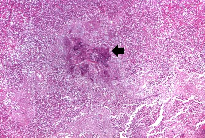
This higher-power photomicrograph shows a central portion of an abscess. Note the absence of any parenchymal lung tissue in this section due to extensive neutrophilic infiltration with liquefaction necrosis of the parenchymal tissue. Masses of leukocytes (primarily neutrophils), fluid ("liquor puris" which is serum, fibrin, etc.), and necrotic debris within an abscess form what is referred to as "purulent material"--or "pus" in lay terminology. The blue-staining mass in the center of this abscess (arrow) represents colonies of bacteria.
File history
Click on a date/time to view the file as it appeared at that time.
| Date/Time | Thumbnail | Dimensions | User | Comment | |
|---|---|---|---|---|---|
| current | 22:23, 18 August 2013 | | 668 × 450 (86 KB) | Seung Park (talk | contribs) | This higher-power photomicrograph shows a central portion of an abscess. Note the absence of any parenchymal lung tissue in this section due to extensive neutrophilic infiltration with liquefaction necrosis of the parenchymal tissue. Masses of leukocyt... |
You cannot overwrite this file.
File usage
The following page uses this file:
