File:IPLab3LobarPneumonia5.jpg
No higher resolution available.
IPLab3LobarPneumonia5.jpg (686 × 450 pixels, file size: 53 KB, MIME type: image/jpeg)
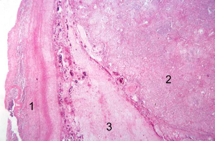
This is a photomicrograph of the interpleural space between the two lung lobules. Thickened pleura with extensive fibrin deposits is present on the left portion of the slide (1) and the right portion of the slide (2) contains the affected lung lobe. Note the extensive fibrin deposits in the interlobar space in the center portion of the slide (3).
File history
Click on a date/time to view the file as it appeared at that time.
| Date/Time | Thumbnail | Dimensions | User | Comment | |
|---|---|---|---|---|---|
| current | 22:17, 18 August 2013 | | 686 × 450 (53 KB) | Seung Park (talk | contribs) | This is a photomicrograph of the interpleural space between the two lung lobules. Thickened pleura with extensive fibrin deposits is present on the left portion of the slide (1) and the right portion of the slide (2) contains the affected lung lobe. No... |
You cannot overwrite this file.
File usage
The following page uses this file:
