File:IPLab2Hypertrophy3.jpg


Size of this preview: 800 × 581 pixels. Other resolution: 1,262 × 916 pixels.
Original file (1,262 × 916 pixels, file size: 163 KB, MIME type: image/jpeg)
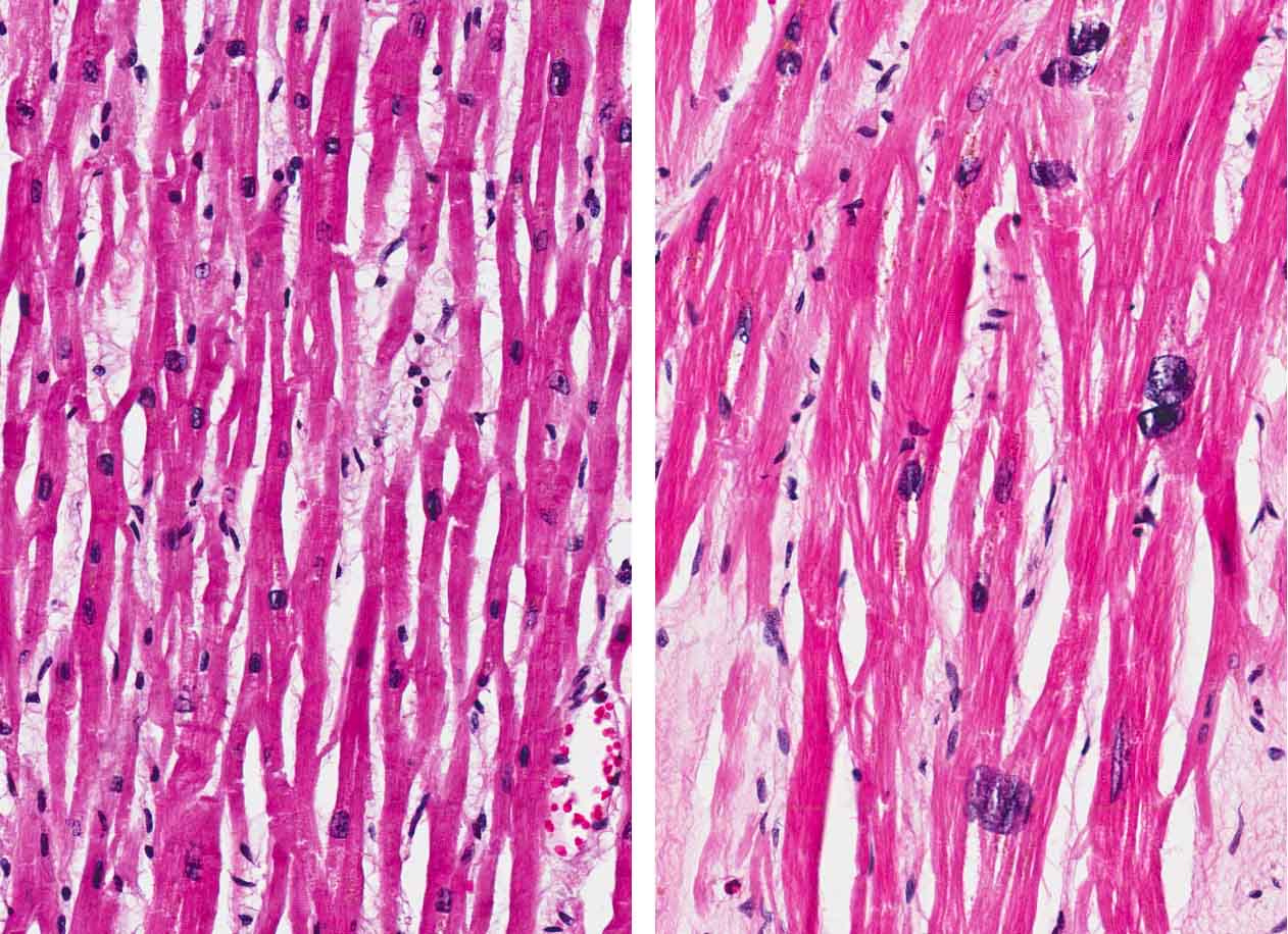
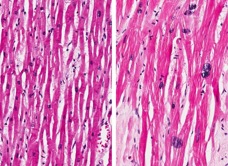
Normal myocardium (left) is compared here to hypertrophied myocardium (right). The muscle fibers are thicker and the nuclei are larger and darker in the hypertrophied myocardium.The clear spaces between the muscle fibers are due to processing artifacts and are not present during life.
File history
Click on a date/time to view the file as it appeared at that time.
| Date/Time | Thumbnail | Dimensions | User | Comment | |
|---|---|---|---|---|---|
| current | 17:26, 4 September 2013 | | 1,262 × 916 (163 KB) | Peter Anderson (talk | contribs) | |
| 18:29, 18 August 2013 | 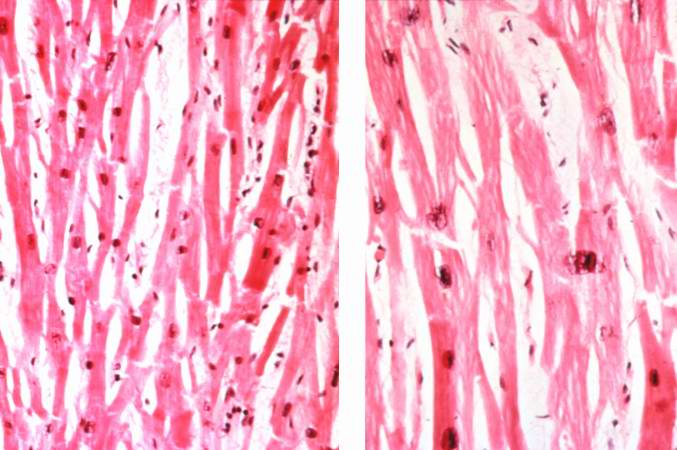 | 677 × 450 (49 KB) | Seung Park (talk | contribs) | Normal myocardium (left) is compared here to hypertrophied myocardium (right). The muscle fibers are thicker and the nuclei are larger and darker in the hypertrophied myocardium.The clear spaces between the muscle fibers are due to processing artifacts... |
You cannot overwrite this file.
File usage
The following page uses this file:
