File:IPLab13CF9.jpg
Revision as of 06:01, 21 August 2013 by Seung Park (talk | contribs) (A higher-power photomicrograph shows the bottom of the intestinal crypts and the other normal layers of the intestine. Even at this magnification, accumulations of eosinophilic debris can be seen in many of the intestinal crypts (arrows).)
No higher resolution available.
IPLab13CF9.jpg (676 × 450 pixels, file size: 87 KB, MIME type: image/jpeg)
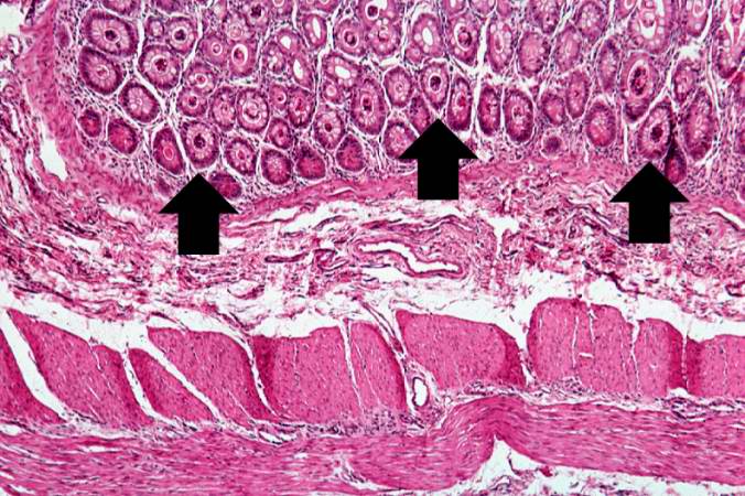
A higher-power photomicrograph shows the bottom of the intestinal crypts and the other normal layers of the intestine. Even at this magnification, accumulations of eosinophilic debris can be seen in many of the intestinal crypts (arrows).
File history
Click on a date/time to view the file as it appeared at that time.
| Date/Time | Thumbnail | Dimensions | User | Comment | |
|---|---|---|---|---|---|
| current | 06:01, 21 August 2013 | | 676 × 450 (87 KB) | Seung Park (talk | contribs) | A higher-power photomicrograph shows the bottom of the intestinal crypts and the other normal layers of the intestine. Even at this magnification, accumulations of eosinophilic debris can be seen in many of the intestinal crypts (arrows). |
- You cannot overwrite this file.
File usage
The following page links to this file: