File:IPLab13CF8.jpg
Revision as of 06:00, 21 August 2013 by Seung Park (talk | contribs) (This low-power photomicrograph of intestine shows the normal layers of the intestine, including the serosa (1), the muscularis (2), the submucosa (3), and the mucosal layer (4) with its deep mucosal crypts. There is yet another cystic space within the ...)
No higher resolution available.
IPLab13CF8.jpg (689 × 450 pixels, file size: 47 KB, MIME type: image/jpeg)
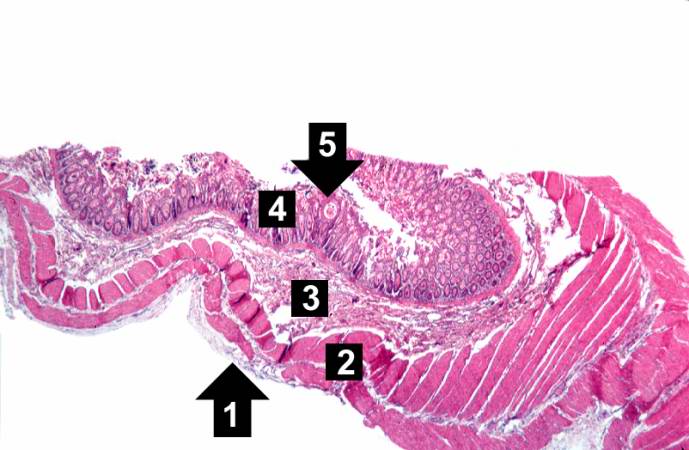
This low-power photomicrograph of intestine shows the normal layers of the intestine, including the serosa (1), the muscularis (2), the submucosa (3), and the mucosal layer (4) with its deep mucosal crypts. There is yet another cystic space within the mucosa (5).
File history
Click on a date/time to view the file as it appeared at that time.
| Date/Time | Thumbnail | Dimensions | User | Comment | |
|---|---|---|---|---|---|
| current | 06:00, 21 August 2013 | | 689 × 450 (47 KB) | Seung Park (talk | contribs) | This low-power photomicrograph of intestine shows the normal layers of the intestine, including the serosa (1), the muscularis (2), the submucosa (3), and the mucosal layer (4) with its deep mucosal crypts. There is yet another cystic space within the ... |
- You cannot overwrite this file.
File usage
The following page links to this file: