File:IPLab11Chagas6.jpg
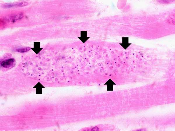
Revision as of 05:08, 21 August 2013 by Seung Park (talk | contribs) (This is a higher-power photomicrograph of an H & E stained heart biopsy from this patient. Note the T. cruzi amastigotes (arrows) within this longitudinal section of a myocyte.)
No higher resolution available.
IPLab11Chagas6.jpg (600 × 450 pixels, file size: 34 KB, MIME type: image/jpeg)
This is a higher-power photomicrograph of an H & E stained heart biopsy from this patient. Note the T. cruzi amastigotes (arrows) within this longitudinal section of a myocyte.
File history
Click on a date/time to view the file as it appeared at that time.
| Date/Time | Thumbnail | Dimensions | User | Comment | |
|---|---|---|---|---|---|
| current | 05:08, 21 August 2013 | | 600 × 450 (34 KB) | Seung Park (talk | contribs) | This is a higher-power photomicrograph of an H & E stained heart biopsy from this patient. Note the T. cruzi amastigotes (arrows) within this longitudinal section of a myocyte. |
- You cannot overwrite this file.
File usage
The following page links to this file: