File:IPLab11Malaria6.jpg
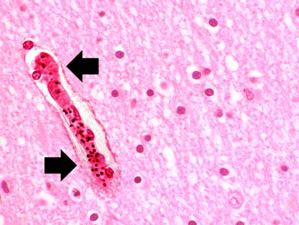
Revision as of 04:55, 21 August 2013 by Seung Park (talk | contribs) (This high photomicrograph was taken from another patient who died of malignant cerebral malaria caused by P. falciparum. In this photomicrograph, a small artery (arrow) can be seen that is full of parasitized RBCs. These RBCs tend to clog small blood v...)
No higher resolution available.
IPLab11Malaria6.jpg (598 × 450 pixels, file size: 47 KB, MIME type: image/jpeg)
This high photomicrograph was taken from another patient who died of malignant cerebral malaria caused by P. falciparum. In this photomicrograph, a small artery (arrow) can be seen that is full of parasitized RBCs. These RBCs tend to clog small blood vessels and lead to cerebral ischemia/hypoxia.
File history
Click on a date/time to view the file as it appeared at that time.
| Date/Time | Thumbnail | Dimensions | User | Comment | |
|---|---|---|---|---|---|
| current | 04:55, 21 August 2013 | | 598 × 450 (47 KB) | Seung Park (talk | contribs) | This high photomicrograph was taken from another patient who died of malignant cerebral malaria caused by P. falciparum. In this photomicrograph, a small artery (arrow) can be seen that is full of parasitized RBCs. These RBCs tend to clog small blood v... |
- You cannot overwrite this file.
File usage
The following page links to this file: