File:IPLab11Malaria2.jpg
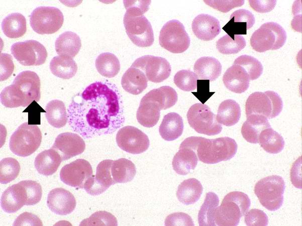
Revision as of 04:54, 21 August 2013 by Seung Park (talk | contribs) (This is another high power photomicrograph of a thin smear of blood from this patient. There is a single eosinophil in this smear along with several RBCs containing ring stage trophozoites (arrows).)
No higher resolution available.
IPLab11Malaria2.jpg (601 × 450 pixels, file size: 36 KB, MIME type: image/jpeg)
This is another high power photomicrograph of a thin smear of blood from this patient. There is a single eosinophil in this smear along with several RBCs containing ring stage trophozoites (arrows).
File history
Click on a date/time to view the file as it appeared at that time.
| Date/Time | Thumbnail | Dimensions | User | Comment | |
|---|---|---|---|---|---|
| current | 04:54, 21 August 2013 | | 601 × 450 (36 KB) | Seung Park (talk | contribs) | This is another high power photomicrograph of a thin smear of blood from this patient. There is a single eosinophil in this smear along with several RBCs containing ring stage trophozoites (arrows). |
- You cannot overwrite this file.
File usage
The following page links to this file: