File:IPLab9Actinomycosis3.jpg
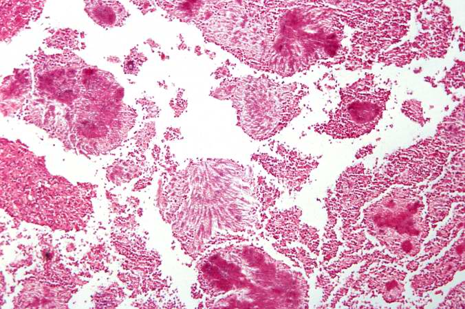
Revision as of 03:59, 21 August 2013 by Seung Park (talk | contribs) (This is a higher-power photomicrograph of actinomycotic colonies in the abscess.)
No higher resolution available.
IPLab9Actinomycosis3.jpg (676 × 450 pixels, file size: 86 KB, MIME type: image/jpeg)
This is a higher-power photomicrograph of actinomycotic colonies in the abscess.
An abscess is a collection of pus (white blood cells) within a cavity formed by disintegrated tissue.
File history
Click on a date/time to view the file as it appeared at that time.
| Date/Time | Thumbnail | Dimensions | User | Comment | |
|---|---|---|---|---|---|
| current | 03:59, 21 August 2013 | | 676 × 450 (86 KB) | Seung Park (talk | contribs) | This is a higher-power photomicrograph of actinomycotic colonies in the abscess. |
- You cannot overwrite this file.
File usage
The following page links to this file: