File:IPLab9Actinomycosis1.jpg
Revision as of 03:59, 21 August 2013 by Seung Park (talk | contribs) (This is a low-power photomicrograph of the retroperitoneal abscess. At this magnification, multiple dark-staining foci can be appreciated. These foci are Actinomyces colonies (arrows). These colonies are known as "sulfur granules" because in gross spec...)
No higher resolution available.
IPLab9Actinomycosis1.jpg (661 × 450 pixels, file size: 39 KB, MIME type: image/jpeg)
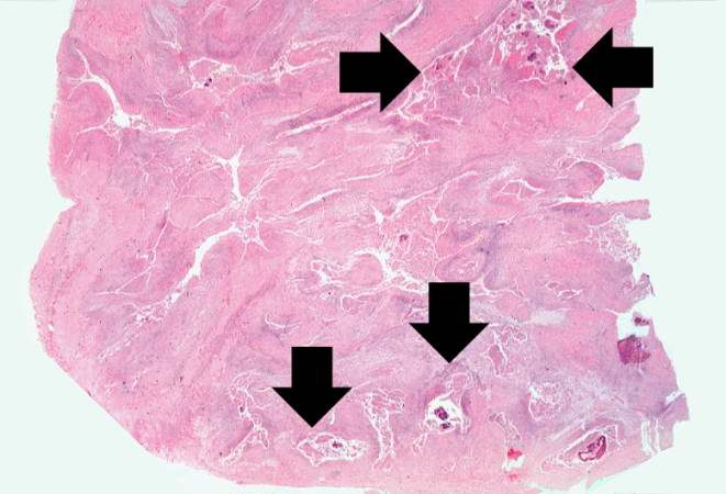
This is a low-power photomicrograph of the retroperitoneal abscess. At this magnification, multiple dark-staining foci can be appreciated. These foci are Actinomyces colonies (arrows). These colonies are known as "sulfur granules" because in gross specimens they are visible to the naked eye as yellow grains, thus resembling grains of sulfur.
An abscess is a collection of pus (white blood cells) within a cavity formed by disintegrated tissue.
File history
Click on a date/time to view the file as it appeared at that time.
| Date/Time | Thumbnail | Dimensions | User | Comment | |
|---|---|---|---|---|---|
| current | 03:59, 21 August 2013 | | 661 × 450 (39 KB) | Seung Park (talk | contribs) | This is a low-power photomicrograph of the retroperitoneal abscess. At this magnification, multiple dark-staining foci can be appreciated. These foci are Actinomyces colonies (arrows). These colonies are known as "sulfur granules" because in gross spec... |
- You cannot overwrite this file.
File usage
The following page links to this file: