File:IPLab9Clostridium6.jpg
No higher resolution available.
IPLab9Clostridium6.jpg (692 × 450 pixels, file size: 55 KB, MIME type: image/jpeg)
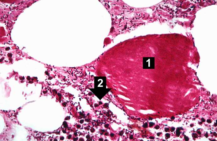
This higher-power photomicrograph of the previous image provides a clearer view of gas bubbles in the tissue, the necrotic hypereosinophilic muscle cell (1), and the mild inflammatory reaction (2). At this magnification, the bacteria located throughout this section can be better appreciated.
File history
Click on a date/time to view the file as it appeared at that time.
| Date/Time | Thumbnail | Dimensions | User | Comment | |
|---|---|---|---|---|---|
| current | 22:57, 20 August 2013 | | 692 × 450 (55 KB) | Seung Park (talk | contribs) | This higher-power photomicrograph of the previous image provides a clearer view of gas bubbles in the tissue, the necrotic hypereosinophilic muscle cell (1), and the mild inflammatory reaction (2). At this magnification, the bacteria located throughout... |
You cannot overwrite this file.
File usage
The following page uses this file:
