IPLab:Lab 11:Schistosomiasis
Clinical Summary
This 30-year-old white male was admitted to the hospital following an upper gastrointestinal hemorrhage with repeated hematemesis. On admission he was tachycardic and had orthostatic hypotension. Endoscopy revealed the presence of esophageal varices. Following fluid replacement and control of bleeding, the patient appeared well. Careful physical exam revealed a palpable spleen extending to the umbilicus. Ascites was not present. The hematocrit was 27%, the white count 12,000 with 30% eosinophils, and the platelet count 98,000. The patient denied alcohol use and had no history of hepatitis. However it was found that two years prior to admission he had worked as a Peace Corps volunteer on a fish-farming project in Ghana. He had been generally healthy while in Africa. He could recall only a fever and cough lasting two or three days, and a brief episode of fever, chills, and headache which was empirically treated as malaria, although no parasites were seen on a single peripheral smear at the time. Since his return to the U.S. eighteen months ago, he had been in good health, but had occasionally noticed some vague left upper quadrant discomfort. Examination of the stool for ova and parasites revealed a few Schistosoma mansoni eggs.
Images
-
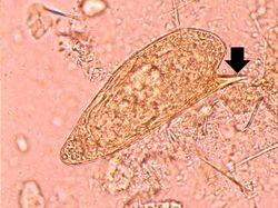 This is a high-power photomicrograph of the patient's fecal specimen containing the characteristic egg of Schistosoma mansoni. Note the spine on the egg (arrow).
 -
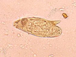 This is high-power photomicrograph of the patient's fecal specimen containing another Schistosoma mansoni egg.
 -
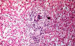 This is a photomicrograph of the liver from another patient with Schistosoma mansoni. These eggs have elicited a robust inflammatory response, including a multinucleated giant cell. These lesions go on to heal by fibrosis resulting in cirrhosis and the classic "pipe-stem" fibrosis.


