Cytologically Yours: CoW: 20131202
Clinical Summary
The patient is an 60 year old male with a remote history of an abdominal melanoma that was excised with negative margins. The patient has been experiencing lower back pain for the past several months and has received epidural injections. As a part of the workup, the patient had a CT which revealed retroperitoneal lymphadenopathy. A CT guided fine needle aspiration and biopsy of a paracaval lymph node was performed.
Past Medical History
- 2003 Melanoma
- Diabetes
- Hypertension
Past Surgical History
- 2013 Arthroscopic knee surgery
- 2003 Excision of melanoma
- 2002 Discectomy
Clinical Plan
The differential diagnosis for otherwise asymptomatic lymphadenopathy in this patient is melanoma, lymphoma, or occult malignancy.
Radiology
- PET CT showed hypermetabolic activity with an SUV of 12.7.
- CT of abdomen and pelvis showed adenopathy adjacent to the aorta and inferior to the vena cava at the level of the right kidney. The largest node measured 4 cm in greatest dimension.
CT


Pathology
Cytology
-
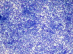 10x magnification of paracaval lymph node. There is a polymorphic lymphoid population with scattered large atypical cells.
 -
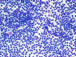 20x magnification of paracaval lymph node. There are small lymphocytes with background lymphoglandular bodies. Scattered eosinophils and large atypical cells with prominent nucleoli.
 -
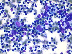 40x magnification of paracaval lymph node. There are atypical binucleated cells among the large atypical cells.


Resident Questions
Final Diagnosis
Cytology
- Positive for malignancy, the differential diagnosis includes melanoma and Hodgkin lymphoma.
Biopsy
- Classical Hodgkin lymphoma, favor mixed type.
Case Discussion
This is a classic case of prostatic adenocarcinoma, metastatic to the spine.