File:IPLab1Tuberculosis3.jpg


No higher resolution available.
IPLab1Tuberculosis3.jpg (676 × 450 pixels, file size: 42 KB, MIME type: image/jpeg)
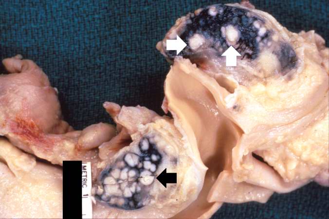
This gross photograph shows hilar lymph nodes from another patient with disseminated tuberculosis. The white, cheesy-appearing nodules (arrows) in the lymph nodes give rise to the descriptive terminology of caseous necrosis. The black pigment in the lymph nodes is anthracotic pigment that has drained from the lungs.
File history
Click on a date/time to view the file as it appeared at that time.
| Date/Time | Thumbnail | Dimensions | User | Comment | |
|---|---|---|---|---|---|
| current | 21:49, 15 August 2013 | | 676 × 450 (42 KB) | Seung Park (talk | contribs) |
You cannot overwrite this file.
File usage
The following page uses this file:
