IPLab:Lab 13:Hyaline Membrane Disease
Clinical Summary
This 28-hour-old white male was the product of an uncomplicated vaginal delivery by a G1 24-year-old mother whose pregnancy had also been uncomplicated. The infant was born pre-term at 36 weeks gestation and was appropriate for gestational age, weighing 2550 grams. Three hours after birth, the infant developed cyanosis and required 40% oxygen to maintain a pink color. Because of a worsening respiratory distress syndrome, the infant (1 day old) was transferred to a tertiary care hospital with a neonatal intensive care unit. Following the transfer, the respiratory distress progressed and the admitting arterial blood gases were pH 7.31, pCO2 35, and pO2 35. The condition of the patient continued to deteriorate despite therapy, and he became "shocky" with efforts to increase blood volume. He developed a persistent bradycardia and died about 4 hours after admission.
Autopsy Findings
The organs generally showed no abnormalities other than those of immaturity expected at this gestational age. There was moderate diffuse subarachnoid hemorrhage and a small amount of blood in the pleural and pericardial cavities.
Images
-
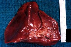 This is a gross photograph of lung demonstrating hyaline membrane disease and atelectasis.
 -
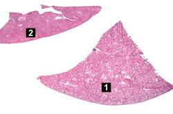 This is a low-power photomicrograph of a triangular-shaped section of lung (1) and an oblong section of liver (2). The lack of open air spaces in this neonatal lung indicates its immaturity.
 -
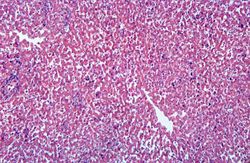 This is a low-power photomicrograph of liver which contains dark blue-stained cells in the hepatic sinusoids. These are immature blood cell precursors and this represents extramedullary hematopoiesis of the liver.
 -
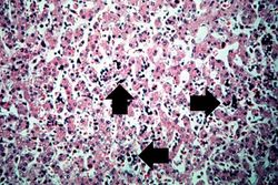 This high-power photomicrograph of liver shows more clearly the immature blood cell precursors (arrows) which represent extramedullary hematopoiesis of the liver. The liver is a normal site of fetal hematopoiesis and, for this stage of gestation, extramedullary hematopoiesis of the liver is normal.
 -
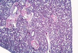 This low-power photomicrograph of lung demonstrates hypercellular pulmonary interstitium and small air spaces (as compared to adult lungs).
 -
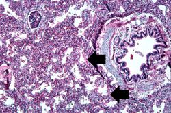 This is a medium-power photomicrograph showing a large bronchus with cartilage. Interstitial congestion with numerous red cells is apparent. Even at this magnification hyaline membranes (arrows) can be seen lining the alveoli.
 -
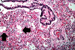 This high-power photomicrograph shows an airway with adjacent lung tissue. Some alveoli have hyaline membranes (arrows). There is severe congestion of the interstitium throughout this section.
 -
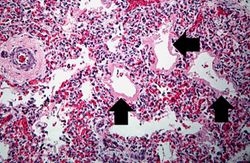 This medium-power photomicrograph shows the pink acellular homogeneous material lining the alveoli which comprises the hyaline membranes (arrows). The interstitium shows congestion, as in previous sections.
 -
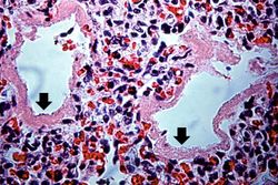 This higher-power photomicrograph shows more clearly the hyaline membranes (arrows) and the congestion in the interstitium.


