IPLab:Lab 6:Hashimoto's Thyroiditis: Difference between revisions
Seung Park (talk | contribs) No edit summary |
|||
| (6 intermediate revisions by 2 users not shown) | |||
| Line 1: | Line 1: | ||
== Clinical Summary == | == Clinical Summary == | ||
This was a 49-year-old woman who complained | This was a 49-year-old woman who complained of tiredness and difficulty concentrating. She had gained weight over the last year and despite warm weather, she felt chilled without a sweater. Family history was significant for hypothyroidism in her mother and older sister. | ||
On physical examination she had an enlarged thyroid gland with a firm, bosselated texture. Serum TSH was markedly elevated and antithyroid peroxidase antibodies were positive. These results supported the clinical impression of hypothyroidism; also, the texture of her thyroid gland and a positive family history suggested an autoimmune etiological factor. She was referred to an endocrinologist; however, before beginning treatment she died suddenly from a ruptured berry aneurysm. | |||
== Images == | == Images == | ||
<gallery heights="250px" widths="250px"> | <gallery heights="250px" widths="250px"> | ||
File:IPLab6Hashimoto1.jpg|This is a gross photograph of thyroid gland taken at autopsy. The gland is only slightly enlarged and has a firm texture. | File:IPLab6Hashimoto1.jpg|This is a gross photograph of thyroid gland taken at autopsy. The gland is only slightly enlarged and has a firm texture. | ||
File: | File:IPLab6Hashimoto3.jpg|This is a low-power photomicrograph of thyroid from this case. Note the large number of blue-staining inflammatory cells in this tissue. These blue cells appear to be forming germinal centers. Some residual thyroid gland tissue can be seen in this section (arrows). | ||
File:IPLab6Hashimoto5b.JPG|This is a higher-power photomicrograph of thyroid from this case showing the inflammatory cells and the residual thyroid tissue (arrow). | |||
File: | |||
File:IPLab6Hashimoto7.jpg|This is a high-power photomicrograph showing the inflammatory cells infiltrating into the residual thyroid tissue (arrows). | File:IPLab6Hashimoto7.jpg|This is a high-power photomicrograph showing the inflammatory cells infiltrating into the residual thyroid tissue (arrows). | ||
File:IPLab6Hashimoto8.jpg|This is a high-power photomicrograph showing the lymphocytes and plasma cells surrounding the thyroid gland epithelium. | File:IPLab6Hashimoto8.jpg|This is a high-power photomicrograph showing the lymphocytes and plasma cells surrounding the thyroid gland epithelium. | ||
</gallery> | </gallery> | ||
== Virtual Microscopy == | |||
<peir-vm>IPLab6Hashimoto</peir-vm> | |||
== Study Questions == | == Study Questions == | ||
| Line 35: | Line 33: | ||
=== Images === | === Images === | ||
* [ | * [{{SERVER}}/library/index.php?/tags/15-endocrine/140-thyroid/141-lymphocytic_thyroiditis PEIR Digital Library: Lymphocytic Thyroiditis Images] | ||
* [http://library.med.utah.edu/WebPath/ENDOHTML/ENDOIDX.html WebPath: Endocrine Pathology] | * [http://library.med.utah.edu/WebPath/ENDOHTML/ENDOIDX.html WebPath: Endocrine Pathology] | ||
Latest revision as of 18:31, 8 July 2020
Clinical Summary
This was a 49-year-old woman who complained of tiredness and difficulty concentrating. She had gained weight over the last year and despite warm weather, she felt chilled without a sweater. Family history was significant for hypothyroidism in her mother and older sister.
On physical examination she had an enlarged thyroid gland with a firm, bosselated texture. Serum TSH was markedly elevated and antithyroid peroxidase antibodies were positive. These results supported the clinical impression of hypothyroidism; also, the texture of her thyroid gland and a positive family history suggested an autoimmune etiological factor. She was referred to an endocrinologist; however, before beginning treatment she died suddenly from a ruptured berry aneurysm.
Images
-
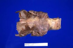 This is a gross photograph of thyroid gland taken at autopsy. The gland is only slightly enlarged and has a firm texture.
 -
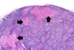 This is a low-power photomicrograph of thyroid from this case. Note the large number of blue-staining inflammatory cells in this tissue. These blue cells appear to be forming germinal centers. Some residual thyroid gland tissue can be seen in this section (arrows).
 -
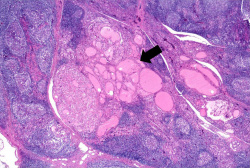 This is a higher-power photomicrograph of thyroid from this case showing the inflammatory cells and the residual thyroid tissue (arrow).
 -
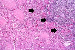 This is a high-power photomicrograph showing the inflammatory cells infiltrating into the residual thyroid tissue (arrows).
 -
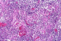 This is a high-power photomicrograph showing the lymphocytes and plasma cells surrounding the thyroid gland epithelium.


Virtual Microscopy
Study Questions
Additional Resources
Reference
- eMedicine Medical Library: Hashimoto Thyroiditis
- eMedicine Medical Library: Hypothyroidism
- Merck Manual: Overview of Thyroid Function
- Merck Manual: Hashimoto's Thyroiditis
- Merck Manual: Hypothyroidism
Journal Articles
- Agrawal P, Ogilvy-Stuart A, Lees C. Intrauterine diagnosis and management of congenital goitrous hypothyroidism. Ultrasound Obstet Gynecol 2002 May;19(5):501-5.