File list
This special page shows all uploaded files.
| Date | Name | Thumbnail | Size | Description | Versions |
|---|---|---|---|---|---|
| 20:29, 16 January 2014 | CytologicallyYoursUnknowns201401-04-10.jpg (file) |  |
730 KB | 3 | |
| 20:29, 16 January 2014 | CytologicallyYoursUnknowns201401-04-09.jpg (file) |  |
613 KB | 2 | |
| 20:28, 16 January 2014 | CytologicallyYoursUnknowns201401-04-08.jpg (file) |  |
679 KB | Reverted to version as of 20:27, 16 January 2014 | 4 |
| 20:27, 16 January 2014 | CytologicallyYoursUnknowns201401-04-07.jpg (file) |  |
525 KB | 2 | |
| 06:06, 21 August 2013 | IPLab13Meningococcemia8.jpg (file) |  |
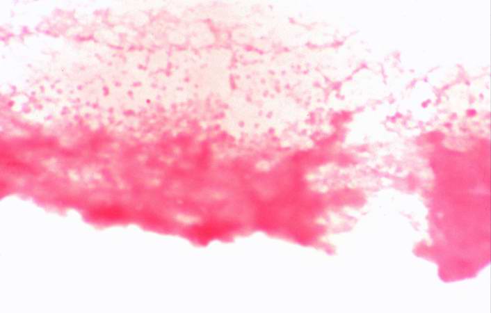
21 KB | This is a higher-power photomicrograph of a smear of cerebrospinal fluid taken at autopsy. Note the Gram-negative cocci in this smear, indicative of N. meningitidis. | 1 |
| 06:05, 21 August 2013 | IPLab13Meningococcemia7.jpg (file) |  |
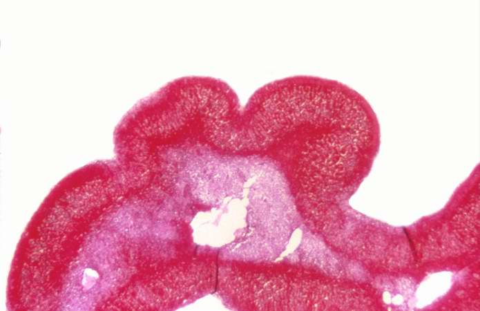
34 KB | This higher-power photomicrograph of the adrenal gland from this case provides an example of hemorrhagic necrosis. | 1 |
| 06:05, 21 August 2013 | IPLab13Meningococcemia6.jpg (file) |  |
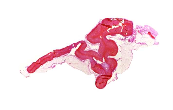
20 KB | This is a low-power photomicrograph of the adrenal gland from this case. Note that the entire gland is hemorrhagic. | 2 |
| 06:05, 21 August 2013 | IPLab13Meningococcemia5.jpg (file) |  |
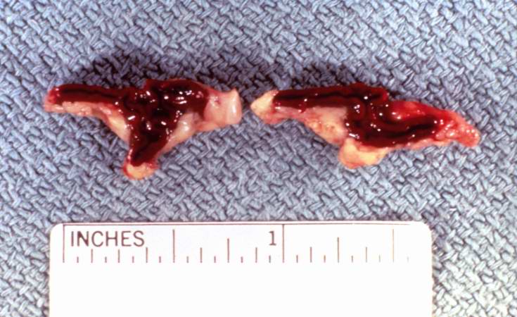
56 KB | This is a gross photograph of cross sections through the adrenal glands from this case. Both adrenal glands are markedly hemorrhagic. | 1 |
| 06:05, 21 August 2013 | IPLab13Meningococcemia4.jpg (file) |  |
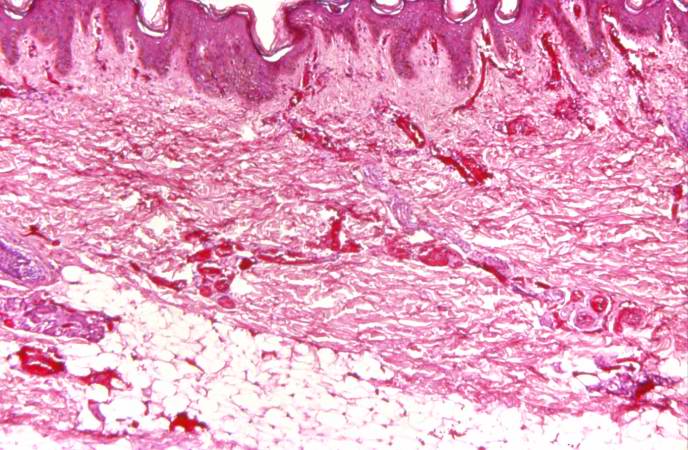
75 KB | This photomicrograph of the skin shows thrombi and fibrin clots in small vessels in the dermis. This is indicative of the endothelial damage caused by the Neisseria meningitidis endotoxin. This endotoxin-induced damage to the endothelium of small blood... | 1 |
| 06:05, 21 August 2013 | IPLab13Meningococcemia3.jpg (file) |  |
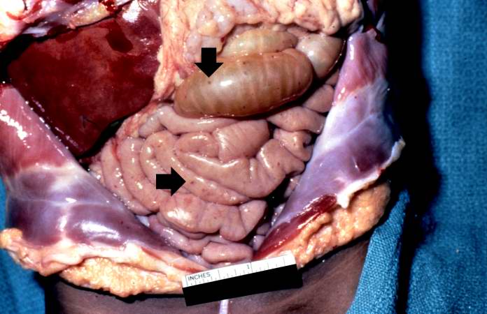
48 KB | In this gross photograph of the abdomen taken at the autopsy, there are petechial hemorrhages on the viscera (arrows). | 1 |
| 06:04, 21 August 2013 | IPLab13Meningococcemia2.jpg (file) |  |
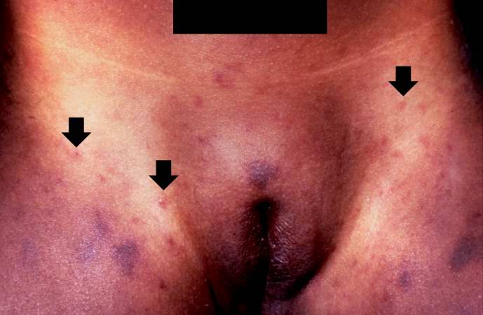
36 KB | This is a closer view of the inguinal region taken at autopsy. The areas of hemorrhage include purpura and petechiae (arrows). | 1 |
| 06:04, 21 August 2013 | IPLab13Meningococcemia1.jpg (file) |  |
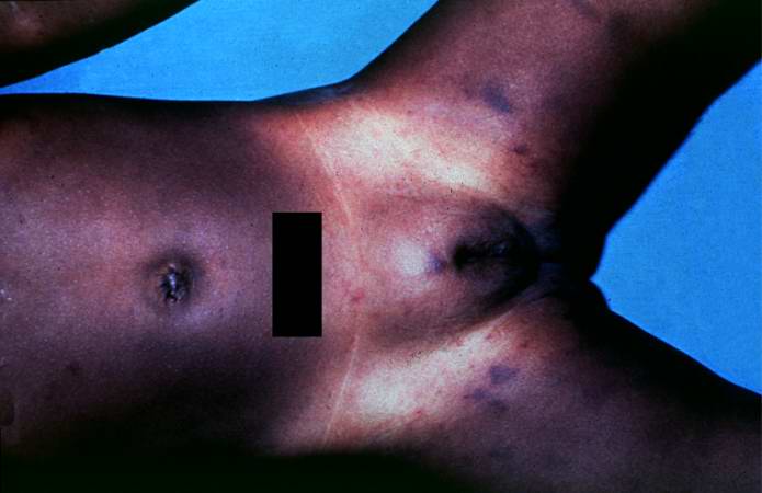
29 KB | In this gross photograph from the autopsy, note the areas of hemorrhage in the inguinal region. | 1 |
| 06:02, 21 August 2013 | IPLab13CF14.jpg (file) |  |
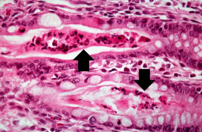
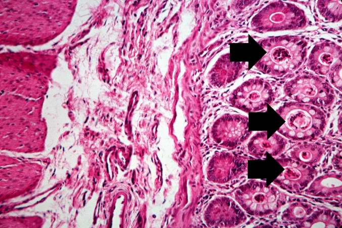
54 KB | Another high-power photomicrograph of intestine shows the vacuolated intestinal epithelial cells lining the crypts and necrotic debris and inspissated secretions within the crypts (arrows). | 1 |
| 06:02, 21 August 2013 | IPLab13CF13.jpg (file) |  |
71 KB | A higher-power photomicrograph of intestine shows the vacuolated intestinal epithelial cells lining the crypts and necrotic debris and inspissated secretions within the crypts (arrows). | 1 |
| 06:01, 21 August 2013 | IPLab13CF12.jpg (file) |  |
86 KB | This is a low-power photomicrograph from another section of the intestine. Saggital sections of the intestinal crypts show the crypts along their full length, extending to the mucosal surface. | 1 |
| 06:01, 21 August 2013 | IPLab13CF11.jpg (file) |  |
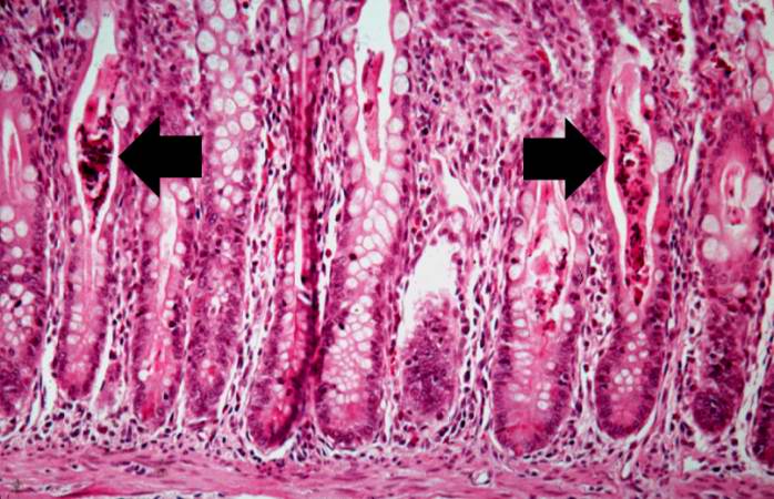
60 KB | This higher-power photomicrograph shows more clearly the eosinophilic debris (arrows) in the intestinal crypts. | 1 |
| 06:01, 21 August 2013 | IPLab13CF10.jpg (file) |  |
74 KB | This is a higher-power photomicrograph showing the eosinophilic debris in many of the intestinal crypts (arrows). | 1 |
| 06:01, 21 August 2013 | IPLab13CF9.jpg (file) |  |
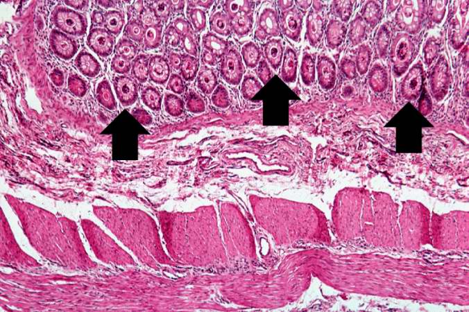
87 KB | A higher-power photomicrograph shows the bottom of the intestinal crypts and the other normal layers of the intestine. Even at this magnification, accumulations of eosinophilic debris can be seen in many of the intestinal crypts (arrows). | 1 |
| 06:00, 21 August 2013 | IPLab13CF8.jpg (file) |  |
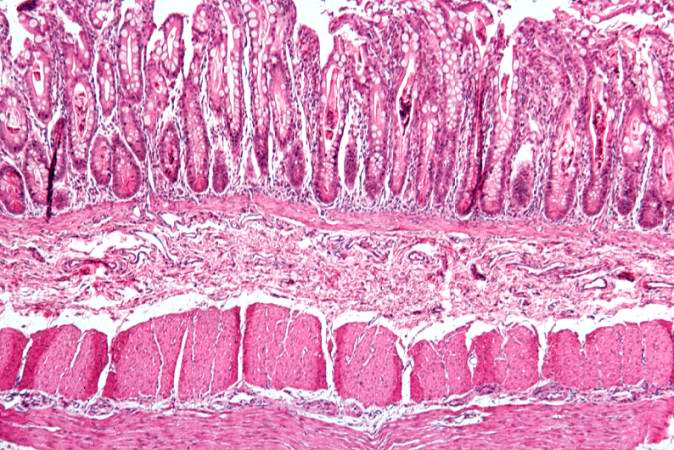
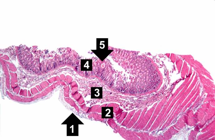
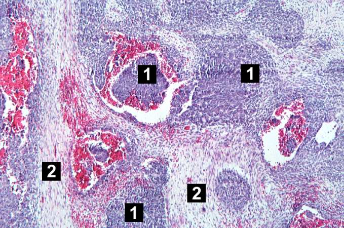
47 KB | This low-power photomicrograph of intestine shows the normal layers of the intestine, including the serosa (1), the muscularis (2), the submucosa (3), and the mucosal layer (4) with its deep mucosal crypts. There is yet another cystic space within the ... | 1 |
| 06:00, 21 August 2013 | IPLab13CF7.jpg (file) |  |
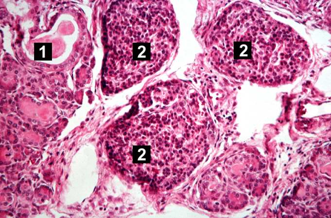
64 KB | This is another high-power photomicrograph showing cystic spaces (1) within the acinar pancreas and a normal islet of Langerhans (2). | 1 |
| 06:00, 21 August 2013 | IPLab13CF6.jpg (file) |  |
73 KB | This high-power photomicrograph shows more clearly these variably-sized cystic spaces within the acinar pancreas. | 1 |
| 06:00, 21 August 2013 | IPLab13CF5.jpg (file) |  |
74 KB | This higher-power photomicrograph shows a cystic space (1) within an acinar lobule. Islets of Langerhans (2) are also visible. | 1 |
| 06:00, 21 August 2013 | IPLab13CF4.jpg (file) |  |
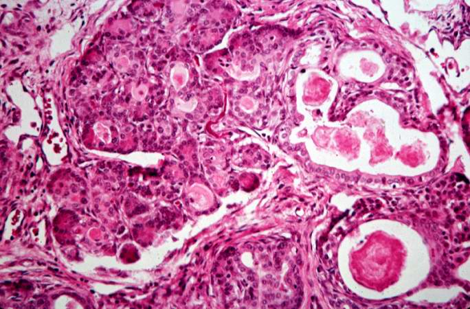
85 KB | This higher-power photomicrograph of the pancreas shows interstitial tissue and the presence of small cystic spaces (1) within the acinar lobules. These spaces are filled with an eosinophilic proteinaceous material. The islets of Langerhans (2) are una... | 1 |
| 05:59, 21 August 2013 | IPLab13CF3.jpg (file) |  |
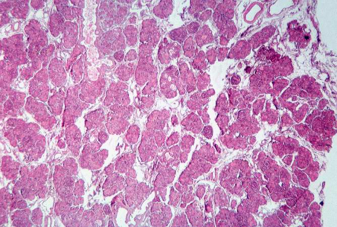
83 KB | This low-power photomicrograph of pancreas shows increased interstitial connective tissue resulting in accentuation of the lobular pattern. | 1 |
| 05:59, 21 August 2013 | IPLab13CF2.jpg (file) |  |
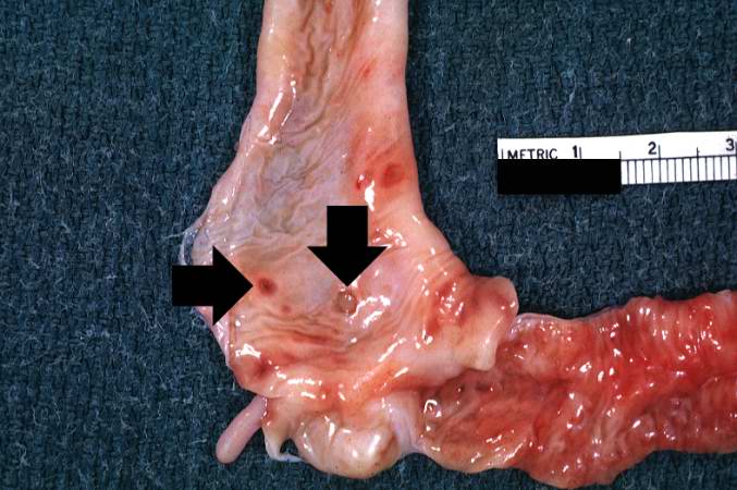
55 KB | This section of duodenum demonstrates dilation, loss of rugae, and areas of ulceration (arrows). | 1 |
| 05:59, 21 August 2013 | IPLab13CF1.jpg (file) |  |
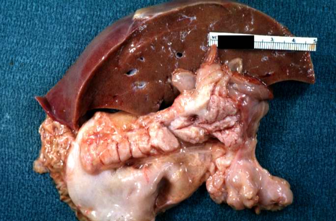
52 KB | This is a gross photograph of liver and pancreas from the autopsy. The pancreas is slightly smaller than normal and it has a mucous consistency. | 1 |
| 05:56, 21 August 2013 | IPLab13WT10.jpg (file) |  |
78 KB | This high-power photomicrograph shows the differences in cell morphology between the blastema (1) and the fibroblast type cells (2). | 1 |
| 05:56, 21 August 2013 | IPLab13WT9.jpg (file) |  |
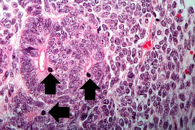
81 KB | This high-power photomicrograph demonstrates tubule formation within the blastema. Note the numerous mitotic figures (arrows). | 1 |
| 05:56, 21 August 2013 | IPLab13WT8.jpg (file) |  |
72 KB | This is a high-power photomicrograph of "tubule" formation within the blastema (arrows). | 1 |
| 05:55, 21 August 2013 | IPLab13WT7.jpg (file) |  |
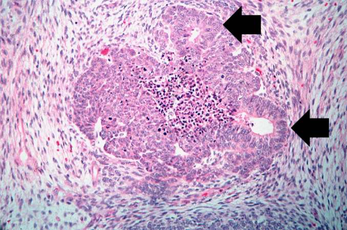
78 KB | This is another medium-power photomicrograph of the tumor. It demonstrates again the two cell types making up this neoplasm. The glands or "tubules" within the blastema are better developed in this section (arrows). | 1 |
| 05:55, 21 August 2013 | IPLab13WT6.jpg (file) |  |
84 KB | This medium-power photomicrograph of tumor shows again the two cell types making up this neoplasm. There are regions within the blastema where the cells form glands or "tubules" (arrows). | 1 |
| 05:55, 21 August 2013 | IPLab13WT5.jpg (file) |  |
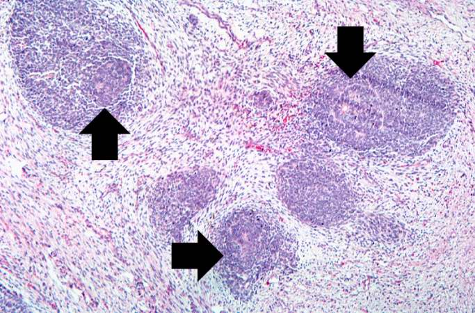
91 KB | This low-power photomicrograph of tumor shows the two cell types making up this neoplasm. The basophilic cellular component termed "blastema" (1) can be distinguished from less cellular eosinophilic areas with fibroblast-like cells (2). | 1 |
| 05:55, 21 August 2013 | IPLab13WT4.jpg (file) |  |
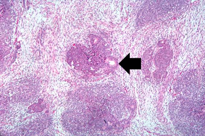
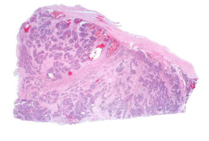
30 KB | This lowest-power view shows the tumor itself; no tissue is present that can be readily identified as normal kidney. There does appear to be a capsule surrounding the tumor. Eosinophilic bands are seen surrounding basophilic islands of cells. These cor... | 1 |
| 05:55, 21 August 2013 | IPLab13WT3.jpg (file) |  |
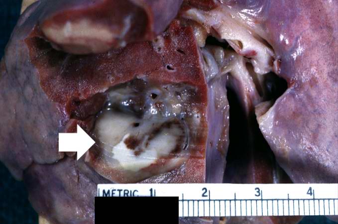
43 KB | This is a gross photograph of lung from this case demonstrating the metastatic tumor nodule (arrow). | 1 |
| 05:54, 21 August 2013 | IPLab13WT2.jpg (file) |  |
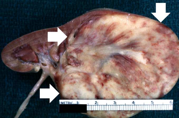
39 KB | This is a closer view of the kidney with Wilms' tumor (arrows). | 1 |
| 05:54, 21 August 2013 | IPLab13WT1.jpg (file) |  |
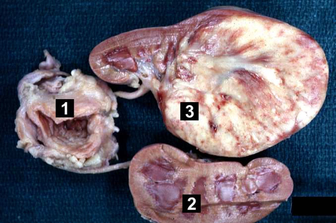
51 KB | This is a gross photograph of a bladder (1) to which are attached a normal kidney (2) and a kidney with Wilms' tumor (3). A large mass extends from the superior pole of the affected kidney. The renal capsule can be seen extending around this tumor. | 1 |
| 05:52, 21 August 2013 | IPLab13Hyaline9.jpg (file) |  |
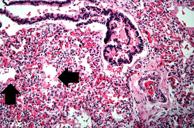
78 KB | This higher-power photomicrograph shows more clearly the hyaline membranes (arrows) and the congestion in the interstitium. | 1 |
| 05:52, 21 August 2013 | IPLab13Hyaline8.jpg (file) |  |
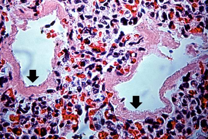
90 KB | This medium-power photomicrograph shows the pink acellular homogeneous material lining the alveoli which comprises the hyaline membranes (arrows). The interstitium shows congestion, as in previous sections. | 1 |
| 05:51, 21 August 2013 | IPLab13Hyaline7.jpg (file) |  |
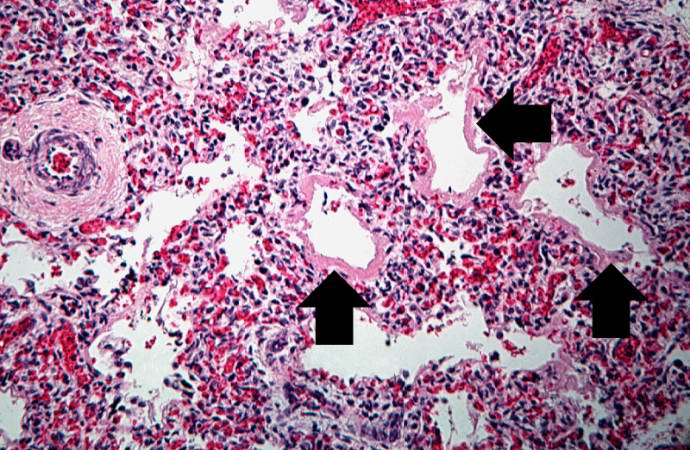
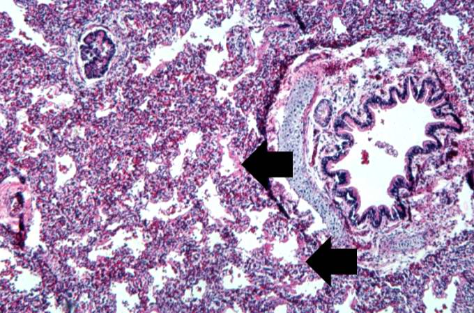
93 KB | This high-power photomicrograph shows an airway with adjacent lung tissue. Some alveoli have hyaline membranes (arrows). There is severe congestion of the interstitium throughout this section. | 1 |
| 05:51, 21 August 2013 | IPLab13Hyaline6.jpg (file) |  |
85 KB | This is a medium-power photomicrograph showing a large bronchus with cartilage. Interstitial congestion with numerous red cells is apparent. Even at this magnification hyaline membranes (arrows) can be seen lining the alveoli. | 1 |
| 05:51, 21 August 2013 | IPLab13Hyaline5.jpg (file) |  |
78 KB | This low-power photomicrograph of lung demonstrates hypercellular pulmonary interstitium and small air spaces (as compared to adult lungs). | 1 |
| 05:51, 21 August 2013 | IPLab13Hyaline4.jpg (file) |  |
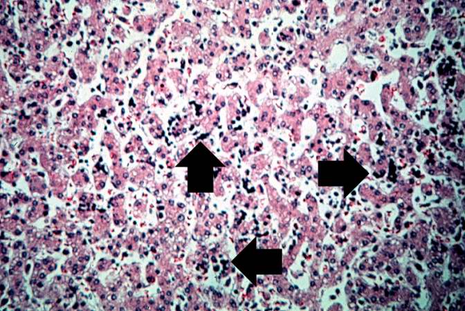
81 KB | This high-power photomicrograph of liver shows more clearly the immature blood cell precursors (arrows) which represent extramedullary hematopoiesis of the liver. The liver is a normal site of fetal hematopoiesis and, for this stage of gestation, extra... | 1 |
| 05:51, 21 August 2013 | IPLab13Hyaline3.jpg (file) |  |
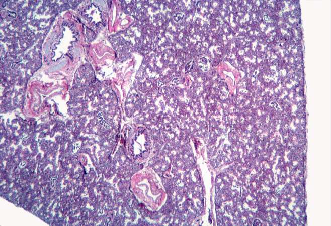
89 KB | This is a low-power photomicrograph of liver which contains dark blue-stained cells in the hepatic sinusoids. These are immature blood cell precursors and this represents extramedullary hematopoiesis of the liver. | 1 |
| 05:50, 21 August 2013 | IPLab13Hyaline2.jpg (file) |  |
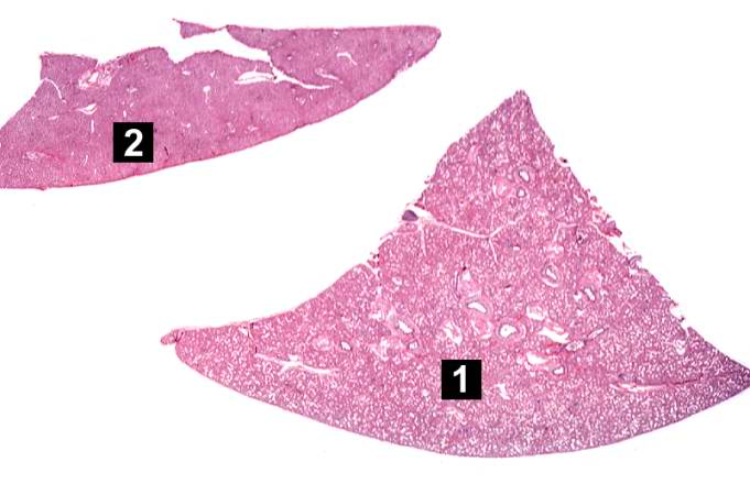
36 KB | This is a low-power photomicrograph of a triangular-shaped section of lung (1) and an oblong section of liver (2). The lack of open air spaces in this neonatal lung indicates its immaturity. | 1 |
| 05:50, 21 August 2013 | IPLab13Hyaline1.jpg (file) |  |
46 KB | This is a gross photograph of lung demonstrating hyaline membrane disease and atelectasis. | 1 |
| 05:48, 21 August 2013 | IPLab13BiliaryAtresia5.jpg (file) |  |
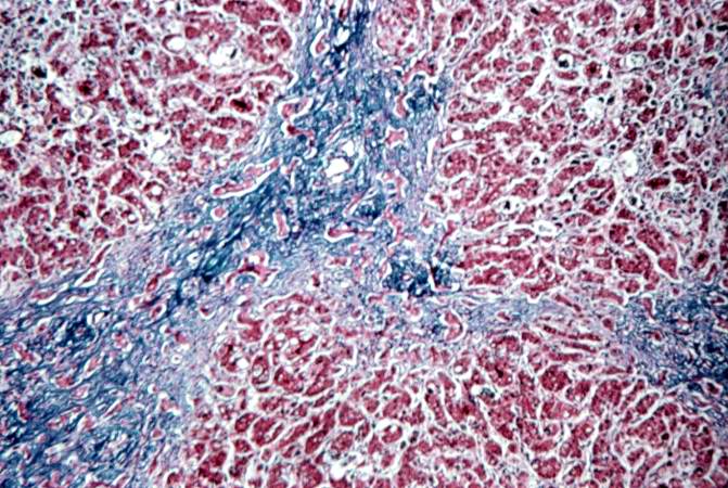
87 KB | This is a medium-power photomicrograph of liver section stained with a trichrome stain to demonstrate the portal fibrosis. The fibrous connective tissue (collagen) stains blue. | 1 |
| 05:48, 21 August 2013 | IPLab13BiliaryAtresia4.jpg (file) |  |
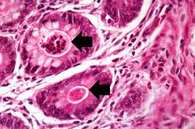
72 KB | This is a high-power photomicrograph of fibrotic portal region with several bile ducts that contain inspissated bile (arrows). Adjacent hepatocytes also contain bile pigments. | 1 |
| 05:48, 21 August 2013 | IPLab13BiliaryAtresia3.jpg (file) |  |
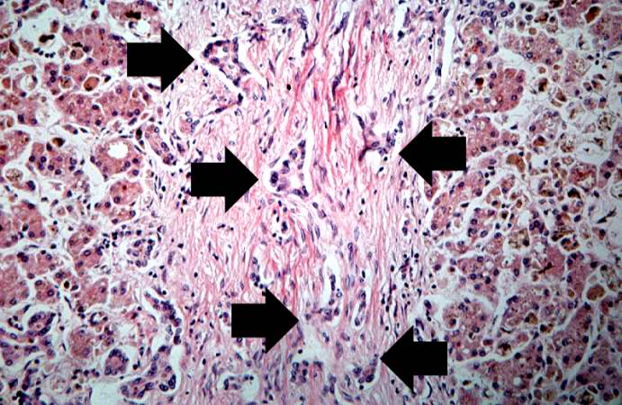
82 KB | This high-power photomicrograph of fibrotic portal region demonstrates proliferation of the bile ducts (arrows). | 1 |
| 05:47, 21 August 2013 | IPLab13BiliaryAtresia2.jpg (file) |  |
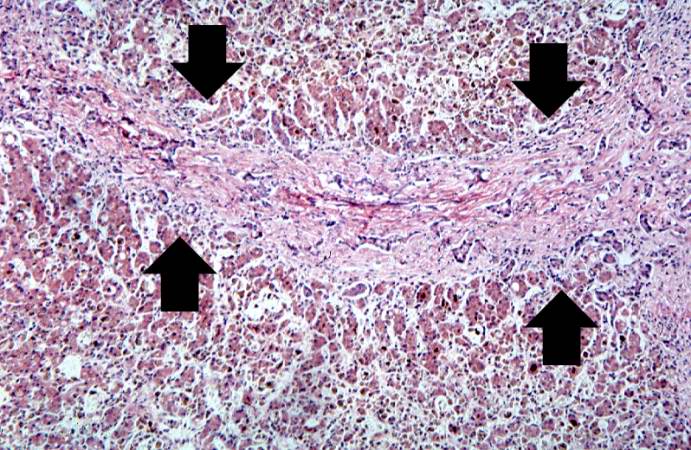
95 KB | This medium-power photomicrograph of liver shows an area of portal fibrosis and bile duct proliferation (arrows). Adjacent to this fibrotic portal region, hepatocytes are seen separated by dilated sinusoids. Throughout this section are found accumulati... | 1 |
| 05:47, 21 August 2013 | IPLab13BiliaryAtresia1.jpg (file) |  |
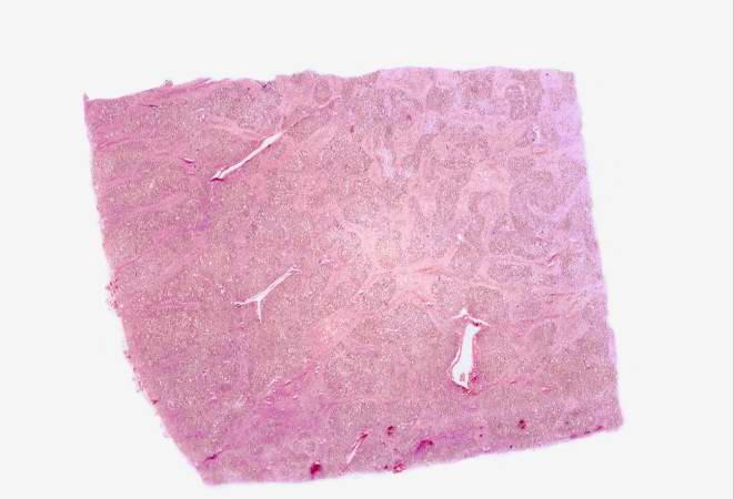
30 KB | This is a low power photomicrograph of a section of liver. Even at this low magnification, areas of fibrosis can be appreciated. | 1 |