Histologic:Chapter 18
Introduction
The microanatomy of the skin varies from one region of the body to another. In studying the slides, compare the similarities as well as differences between skin obtained from different areas of the body.
Slide 46, Thick Skin (H&E)
On this slide of thick skin:
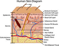
Identify the layers of the epidermis: stratum basale, stratum spinosum, stratum granulosum, stratum lucidum (if present), and stratum corneum.
Stratum basale. This is represented by a single layer of columnar or high cuboidal cells resting on the basement membrane. Note the arrangement and shape of this layer of cells and look for mitoses.
Stratum spinosum. This stratum consists of several layers of polygonal-shaped cells located immediately above the stratum basale. At high power view the delicate processes or spines that project all around the spinous cells and hence the basis for their name: ‘prickle cells.’ These are desmosomes, not intercellular bridges as once believed.
Stratum granulosum. In optimal areas it is seen that this stratum consists of 3-4 layers of squamous-shaped cells containing deeply basophilic granules in the cytoplasm. These are the keratohyalin granules, the first visible indication of the process of keratinization. Recall that the prime purpose of almost all of the cells of the epidermis (with the exception of some of the basal cells) is to become cornified or keratinized. Hence these cells with the keratohyalin granules as well as the spinous cells are referred to as keratinocytes. Another cell type that does not become keratinized is the melanocyte (recognized as the cell with the pale or clear cytoplasm, round dark nucleus, and located in the stratum basale layer). Melanocytes are better seen on slide 4 (thin skin).
Stratum lucidum. Can be seen as a thin light blue or light green layer of cornified cells lying immediately above the stratum granulosum layer.
With high magnification, it can be distinguished from the more basophilic layer of cornified tissue lying above it (stratum corneum).
Stratum corneum. Note the overall thickness of this layer especially when compared with the stratum corneum of thin skin. Also note how much denser it is compared to the loose open weave of the cornified (keratinized) layers of thin skin. Observe the disorganized uppermost layers of the stratum corneum: these are the dead cornified layers being sloughed off (a constant process occurring all over the body surface).
Dermis (Corium, Cutis). On the same slide, study the dermis and note that it is subdivided into the thin papillary layer and the thick reticular layer.
Papillary layer (subepithelial). Observe the fine collagen fibers and some fine elastic fibers in this layer. Note the numerous blood vessels present in the papillae of the papillary layer. Most of these are capillaries forming loops extending up into the primary dermal papillae located between the epidermal ridges; these are “large” capillaries, somewhat wider in diameter than ordinary capillaries. Some of the vascular channels are lymphatic vessels (lymphatic capillaries). A rich lymph system is present in this part of the dermis, but difficult to clearly distinguish from venules or capillaries.
Note an occasional tactile (Meissner’s) corpuscle present in the apical region of a dermal papilla. A Meissner’s corpuscle is an encapsulated sensory nerve ending for light touch (discriminating touch).
Reticular layer. It is named because of the woven arrangement of the collagenous fibers (in the form of a close meshed net, that is, dense irregular connective tissue), not because it is composed of reticular fibers.
Identify blood vessels, nerves, and ducts of sweat glands in the reticular layer. The small veins in thick skin generally have more smooth muscle in their tunica media than comparable veins elsewhere. Secretory units of eccrine sweat glands lie deep in the reticular layer; some are even found in the subcutaneous layer (called the hypodermis, but not part of skin proper). The subcutaneous layer of connective tissue also contains lobules of adipose tissue that are separated by strands of collagenous fibers.
Locate clusters of sections of secretory units of eccrine sweat glands in the dermal-subcutaneous junctional region; each cluster represents one gland. These sweat glands are classified as simple coiled tubular glands. This means that each sweat gland has its own duct and that the secretory portion of the gland is a coiled tube.
In the coiled tubular portion, the secretory cells are simple cuboidal or columnar epithelium and may show pigment or vacuoles in their cytoplasm. Although it may appear that the secretory cells form double layers, all of them touch the basement membrane except where myoepithelial cells intervene. Identify the latter. Only the dark, thin nucleus of the myoepithelial cell can be seen between the basement membrane and the base of the secretory cells. (These myoid or myoepithelial cells are more prominent in apocrine glands.)
Ducts of these sweat glands begin in the coiled portion, then straighten out and pass upward through the dermis. They have a narrow lumen and are lined by two layers of small cells (stratified cuboidal but outer cells may be somewhat rectangular). They lose their epithelial structure as they spiral through the epidermis.
Thin skin: Slides 3, 4, 47, 48
Slide 4, Thin Skin (H&E)
At different magnifications compare thin skin with the sections of thick skin previously studied. Note the extent of the dermis in thin skin (using lowest magnification). One identifying mark of thin skin (aside from the thinner epidermis) is the presence of hair follicles. Only a few are seen on slide 4 (many more are seen on slide 3: scalp).
Which layers of the epidermis are present?
Observe the relatively compact arrangement of the collagen fibers immediately beneath the epidermis and their looser arrangement in the deeper regions of the dermis. No distinctive papillary versus reticular layer of the dermis can be seen.
Note the presence of melanocytes seen among the epithelial cells of the stratum basale. The melanocytes have pale to clear cytoplasm and dark nuclei. It is not possible to see the extent of the melanocytes' cytoplasmic processes that extend up into the spinous layer and surround one to several spinous cells. Pigment is transferred from the melanocytes to the spinous cells.
Slide 3, Scalp (H&E)
From a 28-year old male.
Note the presence of numerous hair follicles on this section of scalp; some sectioned longitudinally show how the stratified squamous epithelium of the epidermis continues as a downward growth to form the epidermal root sheath of the hair follicle. The dermal root sheath is present in favorable sections as an arrangement of the dermis which “follows” the outline of the epidermal root sheath. It is not necessary at this time to study the detailed microscopic anatomy of hair follicles, but be able to recognize sections of hair follicles. Try to identify the layers in cross or oblique sections of follicles.
Observe the presence of numerous sebaceous glands, one or more associated with a hair follicle. Look for sections of erector (arrector pili) muscles (smooth muscle) also associated with hair follicles. The term pilosebaceous unit is given to a hair follicle plus its associated sebaceous gland(s). Note the fine lipid vacuoles in all but the most peripheral small cuboidal cells that rest on the basement membrane of the gland.
The mode of secretion in which the entire cell is lost is called holocrine secretion, and is characteristic of sebaceous glands. Find a duct which connects with the alveolus (also called saccule) of a sebaceous gland. Ducts are lined by stratified squamous epithelium and open into hair follicles; sebum moves up the outside of the hair shaft and reaches the surface of the skin to lubricate it. In some locations (margins of the lips, nipple, glans penis, labia minora) sebaceous glands are not associated with hair follicles, but open directly onto the skin surface.
Study eccrine sweat glands. They are located between hair follicles in the vicinity of the sebaceous glands. Identify their ducts and coiled secretory portions.
Note the extensive hemorrhage in the deep subcutaneous tissue, the result of a skull fracture.
Slide 47, Axillary Skin (H&E)
In addition to the presence of several pilosebaceous units, axillary skin also contains the two types of sudoriferous (sweat) glands: eccrine glands and apocrine glands. Both types of sweat glands are simple coiled tubular glands. The principal structural difference is that secretory portions of apocrine glands are much larger in diameter than the secretory portions of eccrine glands.
Using different magnifications, study the secretory cells of the apocrine sweat glands. These cells are either cuboidal or columnar in shape and the round nucleus with a prominent nucleolus is located in the basilar portion of the cells. Secretory droplets accumulate in the more columnar cells (these droplets are better visualized on slide 48 (PASH) of axillary skin). The ducts are identical to those associated with the eccrine glands but empty into the upper portion of the hair follicle. Where do the eccrine gland ducts empty?
With high magnification study the apical portions of the secretory cells of the apocrine sweat glands. Note how they bulge a considerable way into the lumen. In places they appear to have pinched off and produce the rounded droplets or globules of secretion present in the lumen. In the past, it was assumed that an apical portion of the cell actually was pinched off and discharged (“apocrine secretion”). According to current interpretation, this is probably an erroneous concept resulting from a fixation artifact. Thus, the so-called apocrine sweat gland should really be considered as a modified eccrine or merocrine gland.
Identify the myoepithelial cells in the secretory portions of the apocrine sweat glands; usually only the nucleus can be seen.
Identify eccrine sweat glands. The duct sections are distinct. The secretory cells are pale and vacuolated - a postmortem change.
Identify sections of hair follicles, sebaceous glands, and look for erector pili muscles.
Slide 48, Axillary Skin (PASH)
Note the abundant and prominent PAS-positive secretory material in the apices of apocrine secretory cells. Due to the oblique cut through several apocrine tubules, some of the secretory epithelium is torn away from the basement membrane.
Note, however, the absence of large apocrine tubules such as were seen on slide 47. Instead, smaller tubules are seen lined by tall simple columnar epithelium whose apices are filled with PAS-positive material.
Identify eccrine sweat glands. The ducts are distinct but the secretory cells have marked postmortem degeneration and are sloughing into the lumen.
Mammary Gland
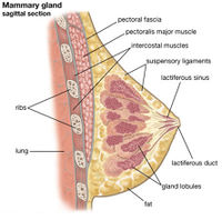
The human mammary gland consists of 15-25 separate compound alveolar glands, each with its own branching duct system. Each of the intralobular ducts of one lobule drains into interlobular ducts, which in turn drain into one of the 15-25 lobar ducts (or lactiferous ducts) that empty separately on the surface of the nipple. The 15 to 25 individual glands or lobes are separated from each other by dense collagenous tissue and abundant adipose tissue, which is primarily responsible for the mass and shape of the breast.
Slide 269, Mammary Gland (H&E)
Slide 269, Mammary gland (H&E) is inactive mammary gland of an adult human female. Histologically, there is a great difference in the mammary gland depending on whether it is inactive, or from a pregnant or lactating individual. Other slides to be studied include inactive gland on slide 268 and two sections from active glands (slides 267 and 270).
Note on slide 269 the large proportion of stroma (dense collagenous connective tissue and fat), the density of the stroma and the amount and distribution of fat.
The stroma is modified around the ducts and ‘embryonic’ alveoli and consists of primitive connective tissue with large fibroblasts, fine fibers, some plasma cells and lymphocytes, abundant ground substance and more numerous blood vessels than elsewhere. This is the intralobular connective tissue.
The interlobular connective tissue has much coarser collagen fibers, fewer fibroblasts, varying numbers of fat cells and fewer vessels.
Study the characteristics of the parenchyma. It consists solely of isolated groups of intralobular ducts and a few interlobular ducts. The ducts are lined with cuboidal or columnar epithelium with myoepithelial cells between its base and the basement membrane.
Slide 268, Mammary Gland (Masson)
Slide 268, Mammary gland (Masson) is tissue from the same inactive gland used to make slide 269. Here, with the Masson’s stain, the differences between intralobular duct connective tissue and interlobular duct connective tissue are more easily distinguishable.
Again note the delicate connective tissue around the intralobular ducts and alveoli. The latter are termed “embryonic” because they resemble the alveoli seen in some infants at term, which are hormonally stimulated to produce “witch’s” milk.
Slide 270, Mammary Gland (H&E)
Slide 270, Mammary gland is from a lactating gland.
Note the tremendously increased proportion of glandular tissue to stroma and the resulting formation of lobes and lobules. Observe the branching of some of the secretory alveoli.
The alveoli have a lumen varying from 50 to 100 μm in diameter lined with simple low cuboidal to low columnar epithelium.
In both the alveolar ducts (intralobular) and the interlobular ducts can be seen stained secretory material (the denatured proteinaceous portion of milk). The same material fills almost all the lumina of the alveoli; the scalloped outline of the secretory material in the lumina of the alveoli is an artifact of the preparation.
Note the large size of some of the intralobular ducts; they are large because they are distended with secretion. This ductal epithelium is simple low cuboidal. Some of the interlobular ducts are smaller than the intralobular (alveolar) ducts.
The interlobular and perilobular stroma is still dense collagenous fibers.
The primitive intralobular stroma has been largely “used up” to provide “room” for the increased numbers of alveoli and ducts, but traces of it remain.
Note the increased number and size of blood vessels in both lobular and interlobular locations.
Slide 267, Mammary Gland (PASH)
Slide 267, Mammary gland is from a lactating gland.
The alveoli are circular and much smaller, on the average, than those seen in slide 270.
Note the cuboidal epithelium lining the alveolar lumina, the vacuolated cytoplasm due to dissolved-out lipid and the PAS-positive material in the alveolar lumina.
The nuclei of the myoepithelial cells are easier to locate on this slide; they appear as elongated, narrow structures located between the epithelial cell and the basement membrane. The cytoplasm of the myoepithelial cells does not stain with either PASH or H&E.
Oxytocin, the milk letdown factor elaborated from the neurohypophysis, is a hormone that stimulates the myoepithelial cell to contract, and in so doing helps squeeze out or eject the milk from the alveoli.
Until evidence from EM studies proved otherwise, the consensus among histologists and physiologists was that milk was produced as an apocrine type of secretion, that is, portions of the alveolar epithelial cell were believed to break off and form part of the secretory product. Current EM information indicates that the protein component of milk is released as a merocrine type of secretion. The process of protein release is called exocytosis.
The lipid component of milk is released from the cell surrounded or enveloped by a membrane derived from the cell’s plasma membrane and an exceeding thin rim of cytoplasm. Technically, this type of secretions is of the apocrine method but the amount of cytoplasm lost is far less than classical histologists would describe for “typical” apocrine gland.”
Nipple Microanatomy
Slide 272, Nipple (Masson's)
Slide 272, Nipple (Masson’s) is a section of nipple and part of the surrounding areolar tissue from a human female breast.
Note the thin epidermal stratified squamous epithelium pierced by numerous “fingers” of dermal papillae. The latter are extremely long and branching particularly in the areolar region around the nipple.
Observe the large sebaceous glands lying within the dermis. Some of these glands are part of hair follicles (pilosebaceous unit); others are free or independent of the hair follicle.
Look for large lactiferous ducts, lined with two-layered stratified cuboidal or low columnar epithelium. Smaller ducts (which lead into the lactiferous duct) may be seen towards one side of the section. These ducts merge directly into alveolar or secreting portions (units) of the mammary gland, which are lined with simple cuboidal to low columnar epithelium.
Numerous bundles of smooth muscles are oriented circumferentially (therefore most are seen cut in x-section) in the nipple at its base and longitudinally around the lactiferous duct. Contraction of the muscle allows the nipple to become erect. Nerve innervation of the nipple is primarily at the tip -not at its sides or in the areola. This is functionally significant because the infant’s sucking stimulus is required for continued normal lactation.
| ||||||||||||||||||||||||||||||||||||||||||||||||||||||||||||||

