Cytologically Yours: CoW: 20140106
Contents
Clinical Summary
64 year old male who presented to the ED with shortness of breath.
Past Medical History
- Hypertension
- Dyslipidemia
- Recent diagnosis of metastatic melanoma of unknown primary
Past Surgical History
- Metastatic melanoma of unknown primary diagnosed in an enlarged axillary mass that was removed several months previously.
Radiology
- CXR and CT showed multiple pulmonary nodules and bilateral pleural effusions.
Clinical Plan
Thoracentesis
Pathology
Cytology
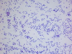
10x magnification of a very cellular specimen. (DQ)
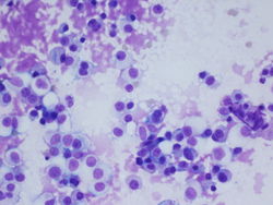
40x magnification showing cells that have ample cytoplasm and prominent nucleoli.(DQ)
40x magnification showing Cells with abundant cytoplasm some with granular material in the cytoplasm. These cells also have prominent nucleoli.
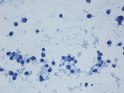
10x magnification of a very cellular specimen. (Pap)
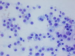
40x magnification of cells with prominent nucleoli and dusty cytoplasm. (Pap)


Final Diagnosis
Cytology
- Metastatic melanoma.
Cell Block
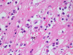
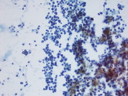
40x magnification of cell block showing cells with pigment in the cytoplasm.
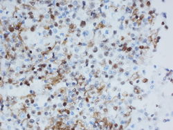
20x magnification of MITF staining of the cell block.


Discussion
Melanoma is "the great mimicer", it's cytomorphology can be very variable. Prominent nucleoli and pigment are some of the features that can help guide a pathologist to the diagnosis. The use of the immunohistochemical markers MART-1, S100, MITF,and MelanA are useful for the diagnosis of melanoma.
| ||||||||