4/7
)
)
)
)
)

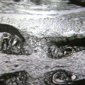
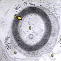
ELECTRON MICROSCOPY: NERVOUS: NERVE: Myelinated peripheral nerve; RCH/AMC1399, small myelinated axon, inner mesaxon is clearly seen, distinct basal lamina at junction of Schwann cell and endoneurium, excess of basal lamina is noted in two locations.
- Author
- Peter Anderson
- Posted on
- Tuesday 6 August 2013
- Tags
- electron microscopy, nerve, nervous
- Albums
- Visits
- 2051
0 comments