89/90
)
)
)
)
)
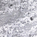
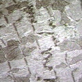
, endoplasmic reticulum; RCH/P&SG3540, cytoplasmic detail of one Purkinje cell showing two Nissl bodies, many of the polyribosomes appear to be free in the intercisternal space, monkey brain. N-Nissl bodies, G-Golgi apparatus, A-small myelinated axons")
ELECTRON MICROSCOPY: CELL: Nissl bodies, Purkinje cell of cerebellum. (EM-labeled), endoplasmic reticulum; RCH/P&SG3540, cytoplasmic detail of one Purkinje cell showing two Nissl bodies, many of the polyribosomes appear to be free in the intercisternal space, monkey brain. N-Nissl bodies, G-Golgi apparatus, A-small myelinated axons
- Author
- Peter Anderson
- Posted on
- Tuesday 6 August 2013
- Tags
- cell, electron microscopy
- Albums
- Visits
- 1824
0 comments