14/17
)
)
)
)
)
. Fat necrosis can calcify in large lesions. (MAMMOGRAM)")
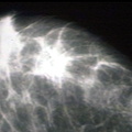
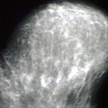
RADIOLOGY: BREAST: Case 2 Left mammogram Dx: Fibroadenoma; Left breast mass in the upper half of breast, close to the chest wall and axilla. This is dense, round and well circumscribed containing 3 or 4 large course calcium deposits (calcification). Fat necrosis can calcify in large lesions. (MAMMOGRAM)
- Author
- Peter Anderson
- Posted on
- Tuesday 6 August 2013
- Tags
- breast, fibroadenoma, radiology
- Albums
- Visits
- 3240
0 comments