)
)
, and glands similar to the male prostatic glands are often also present. Most female diverticula are acquired rather than congenital and occur posteriorly in the midurethra. They are thought to result from infected periurethral glands that form abscess cavities that then rupture into the urethral lumen. Other possible causes of acquired diverticula include trauma during childbirth, instrumenation and fulguration of urethral lesions, urethral strictures or stones. Both benign and malignant tumors arising within the diverticulum itself have also been described in case reports. Acquired diverticula are thought to be present in up to 3% of asymptomatic females. Complications can include stone formation, dysuria, frequent urinary tract infections and dyspareunia, as well as post-void dribbling. Physical exam is usually normal although a palpable, bulging mass is occasionally present and this was the case with the patient presented here in whom one of her diverticular stones had actually begun to erode through the diverticular wall which was clearly visible at her introitus. Other than location, the differentiation between congenital versus acqured diverticula can be very difficult. Congenital diverticula usually occur in the anterior urethra and are almost exclusively a male phenomenon. The appearance of urethral diverticula can be quite variable dependingon the location and the patients sex as well. They can be multiple, or multilocular, saccular, tubular or ovoid and can have a variable number of openings into the urethral lumen. In females, the diverticulum is usually short-necked and tubular or oval. Cystoscopy is not very sensitive, missing up to 60% of cases. In addition to a VCUG, a double-balloon retrograde urethrogram can also be performed, and carefully positioned post-void excretory urogram films can also often correctly diagnose the condition.")
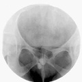
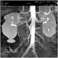
RADIOLOGY: GENITOURINARY: GU: Case# 34660: TWO LARGE CALCULI IN A URETHRAL DIVERTICULUM. A 53 year-old Black female who complained of a sensation of something "dropping" in her pelvis several months prior to imaging as well as dysuria that resolved with oral antibiotics several weeks ago. Cystoscopy done just prior to radiologic evaluation was unremarkable. Two large radiopaque stones projecting over the lower pubic symphysis on scout film. VCUG demonstrated a posterior urethral diverticulum which contained both sontes within it. Bladder was normal. The diverticulum opening was located near the bladder neck. The female urethra, which is homologous to the posterior urethra in the male minus the verumontanum, is approximately 4 cm long. It courses obliquely downward from the internal urethral opening and is slightly curved, although its course is normally quite variable. The urethra contains periuurethral glands (throughout its length?), and glands similar to the male prostatic glands are often also present. Most female diverticula are acquired rather than congenital and occur posteriorly in the midurethra. They are thought to result from infected periurethral glands that form abscess cavities that then rupture into the urethral lumen. Other possible causes of acquired diverticula include trauma during childbirth, instrumenation and fulguration of urethral lesions, urethral strictures or stones. Both benign and malignant tumors arising within the diverticulum itself have also been described in case reports. Acquired diverticula are thought to be present in up to 3% of asymptomatic females. Complications can include stone formation, dysuria, frequent urinary tract infections and dyspareunia, as well as post-void dribbling. Physical exam is usually normal although a palpable, bulging mass is occasionally present and this was the case with the patient presented here in whom one of her diverticular stones had actually begun to erode through the diverticular wall which was clearly visible at her introitus. Other than location, the differentiation between congenital versus acqured diverticula can be very difficult. Congenital diverticula usually occur in the anterior urethra and are almost exclusively a male phenomenon. The appearance of urethral diverticula can be quite variable dependingon the location and the patients sex as well. They can be multiple, or multilocular, saccular, tubular or ovoid and can have a variable number of openings into the urethral lumen. In females, the diverticulum is usually short-necked and tubular or oval. Cystoscopy is not very sensitive, missing up to 60% of cases. In addition to a VCUG, a double-balloon retrograde urethrogram can also be performed, and carefully positioned post-void excretory urogram films can also often correctly diagnose the condition.
- Author
- Peter Anderson
- Posted on
- Thursday 1 August 2013
- Tags
- Genitourinary, radiology
- Albums
- Visits
- 2718
0 comments