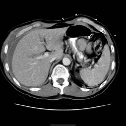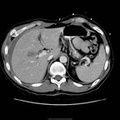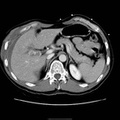
RADIOLOGY: PANCREAS: Case# 47: PANCREATIC TRANSECTION. 32 year old female s/p MVA one day prior now with elevated amylase and lipase. There is a large amount of ascites. There is disruption of the pancreatic head just anterior to the portal vein confluence. Pancreatic transection is uncommon and associated with a high mortality since it is often clinically occult. In complete transection, CT images may show two ends separated by low-density fluid that remains relatively confined to the anterior pararenal space in the immediate post-injury period. Pancreatic enlargement, fluid collections, and lucent defects may also be present. In incomplete transections, tissue displacement may be minimal, and lacerations may be difficult to identify, however, but may be suggested by unexplained thickening of the anterior renal fascia.
- Author
- Peter Anderson
- Posted on
- Thursday 1 August 2013
- Albums
- Visits
- 2096


0 comments