)
)

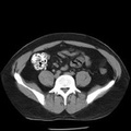
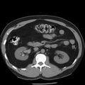
RADIOLOGY: GENITOURINARY: GU: Case# 34075: URETERAL CALCULUS. Patient is a 38 year old male who had CT scan yesterday at Kirklin Clinic for symptoms suggestive of diverticulitis. That CT scan did reveal mild diverticulitis. The patient then had severe right sided flank pain radiating to the groin starting this morning, with hematuria. There is residual contrast within the collecting system on the right with mild hydroureteronephrosis down to the right UVJ. At the right UVJ there is a 1 to 2 mm calcific density in the ureter. There are at least 3 up to 1 to 2 mm calcific densities within the collecting systems centrally on the right and 2 to 3, 1 to 2 mm calcific densities in the central collecting system on the left kidney as well. Colonic diverticulosis and mild changes of diverticulitis again noted. On retrospective review of yesterdays CT scan at wide window levels, the calculus now seen at the right UVJ was present in the upper ureter but there was not obstruction or dilatation at the time.
- Author
- Peter Anderson
- Posted on
- Thursday 1 August 2013
- Tags
- Genitourinary, radiology
- Albums
- Visits
- 1005
0 comments