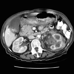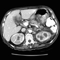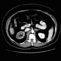
RADIOLOGY: KIDNEY: Case# 33940: XANTHOGRANULOMATOUS PYELONEPHRITIS; CHOLELITHIASIS. 78 year old female with clinical history of gastric outlet obstruction. 1. The left kidney is markedly abnormal with hydronephrosis and perinephric inflammatory change. These findings are most consistent with xanthogranulomatous pyelonephritis. These findings in the left kidney could also be caused by other chronic granulomatous infection such as tuberculosis or fungal infection. 2. The left renal vein is retroaortic. 3. Cholelithiasis. 4. Small bilateral pleural effusions. 5. Stomach wall appears mildly thickened. This is not specific. There is no intra-abdominal adenopathy.
- Author
- Peter Anderson
- Posted on
- Thursday 1 August 2013
- Albums
- Visits
- 1018


0 comments