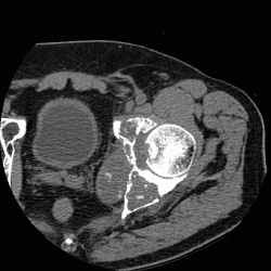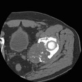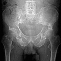
RADIOLOGY: MUSCULOSKELETAL: Case# 33932: CHONDROSARCOMA. Seventy year old male patient with suspicious pathological fracture of the left hip. Large destructive lesion of the left acetabulum with break through of the cortex medially. There is some chondroid matrix of the inferior aspect of the tumor. Chondrosarcoma is the most likely diagnosis. The mass measures approximately 6.0cm in transverse dimension x 6.6cm in the cranial caudad x approximately 6cm from anterior to posterior dimension. The mass replaces the marrow of the quadrilateral plate and the anterior and posterior columns. The mass breaks through the medial cortex of the acetabulum and extends into the pelvic fat where it displaces the internal iliac vessels. There is no invasion of the bladder or rectum.
- Author
- Peter Anderson
- Posted on
- Thursday 1 August 2013
- Tags
- musculoskeletal, radiology
- Albums
- Visits
- 2189


0 comments