)
)

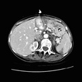
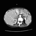
RADIOLOGY: HEPATOBILIARY: Case# 33074: HEPATIC ARTERY PSEUDOANEURYSM. 59 year old female. 1. Two large aneurysm or pseudoaneurysms involving the hepatic arteries are seen. The 6 x 5 cm one in the left hepatic artery has been successfully embolized and there appears to be no flow enhancement. Dilated intrahepatic ducts in the lateral segment of the left lobe are likely secondary to the mass effect within the falciform ligament. A 3 x 3 cm partially thrombosed pseudoaneurysm involving the right hepatic artery is identified. This causes minimal adjacent intrahepatic ductal dilatation involving the posterior segment of the right lobe. There is compression and likely occlusion of the left portal vein as well as the posterior division of the right portal vein. 2. Splenic infarcts. 3. Small amount of ascites. 4. Very large pericardial effusion as well as bilateral large pleural effusions and passive atelectasis of the left lower lobe. 5. Differential diagnosis for these large visceral aneurysms or pseudoaneurysm must include vasculitis including polyarteritis nodosa as well as mycotic origin. Given the large pericardial and pleural effusions SLE would also be considered in the differential diagnosis.
- Author
- Peter Anderson
- Posted on
- Thursday 1 August 2013
- Tags
- hepatobiliary, radiology
- Albums
- Visits
- 762
0 comments