18428/33648
)
)
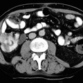
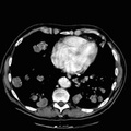
 with relatively high attenuation borders probably representing fat necrosis or inflamatory process of the omentum rather than a neoplasm. 3. Small left pleural effusion with left lower lobe atelectasis. 4. A hiatal hernia is present. 5. Fatty infiltration within the liver with an area of more focal fat infiltration at the porta hepatis.")
RADIOLOGY: GASTROINTESTINAL: GI: Case# 33072: ?OMENTAL FAT NECROSIS & APPENDIX MUCOCELE; S/P TRANSHIATAL ESOPHAGECTOMY. Patient is a 61 year old male with abdominal pain and a palpable mass. 1. No evidence for appendicitis. 2. Irregular predominantly fat attenuation lesions are seen orienting in a longitudinal direction along the anterior abdominal wall (adjacent to the anterior peritoneum) with relatively high attenuation borders probably representing fat necrosis or inflamatory process of the omentum rather than a neoplasm. 3. Small left pleural effusion with left lower lobe atelectasis. 4. A hiatal hernia is present. 5. Fatty infiltration within the liver with an area of more focal fat infiltration at the porta hepatis.
- Author
- Peter Anderson
- Posted on
- Thursday 1 August 2013
- Tags
- gastrointestinal, radiology
- Albums
- Visits
- 666
0 comments