)
)

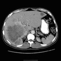
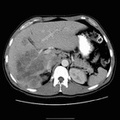
RADIOLOGY: HEPATOBILIARY: Case# 33359: SCLEROSING CHOLANGIOCARCINOMA, ULCERATIVE COLITIS & S/P CHENOEMBOLIZATION/ ? CHOLECYSTITIS. The patient is a 35-year-old male who now presents with right upper quadrant pain and fevers. 1. Large low attenuation, heterogeneous mass in the right lobe of the liver with a smaller similar appearing lesion in the left lobe of the lesion. The most likely consideration in this patient would be cholangiocarcinoma. Other diagnostic considerations would be metastases, presumably from a GI malignancy, though none is identified on this CT scan. Lymphoma can give a similar appearance to the retroperitoneal para-aortic nodes, but the liver lesions would be atypical for lymphoma. These lesions would also be atypical for abscess. 2. Lymphadenopathy as described above. 3. Intrahepatic focal biliary ductal dilatation.
- Author
- Peter Anderson
- Posted on
- Thursday 1 August 2013
- Tags
- hepatobiliary, radiology
- Albums
- Visits
- 685
0 comments