18611/33648
)
)

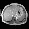
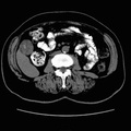
RADIOLOGY: HEPATOBILIARY: Case# 33343: GB CANCER. The patient is a 73 year old female. 1. An ill-defined enhancing soft tissue mass is seen extending from the gallbladder wall into the lumen of the gallbladder. This lesion is highly suspicious for gallbladder carcinoma. There is no evidence for local extension or metastatic disease. 2. Diffuse fatty infiltration of the liver. 3. Sigmoid diverticulosis. 4. Cyst in the intrapolar region of the right kidney.
- Author
- Peter Anderson
- Posted on
- Thursday 1 August 2013
- Tags
- hepatobiliary, radiology
- Albums
- Visits
- 857
0 comments