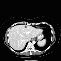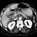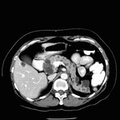
RADIOLOGY: KIDNEY: Case# 33009: VON HIPPEL-LINDAU DISEASE. This is a 44 year old white female status post partial right nephrectomy and left total nephrectomy for renal cell cancer. Comparison is made with a prior CT. The liver is normal in size and contains multiple small hepatic cysts. The left kidney is surgically absent. Multiple simple renal cysts are noted in the right kidney. A complex septated cyst near the upper pole of the right kidney has slightly increased in size. Pre and post-contrast evaluation of the cyst measures 18 and 25 Hounsfields units respectively. However, at the inferior margin, it appears to enhance more. Multiple pancreatic cystic lesions are again noted. The largest of these contains multiple septations and is located in the region of the head of the pancreas. This may represent a serous cystadenoma. von Hippel-Lindau disease is a rare autosomal dominant disorder with variable expressivity characterized by a variety of benign and malignant neoplasms widely dispersed throughout the body, the most characteristic being retinal angiomas and cerebellar hemangioblastomas. Abdominal neoplasms include multiple renal and pancreatic cysts, small renal adenomas, and frequent multiple and bilateral renal adenocarcinomas. These are best visualized using CT. Although CNS abnormalities usually predominate, renal cell carcinoma is a major cause of death. Renal carcinomas may be small and occur within the cysts making early detection difficult. Patients and their relatives should be screened periodically using CT.
- Author
- Peter Anderson
- Posted on
- Thursday 1 August 2013
- Albums
- Visits
- 904


0 comments