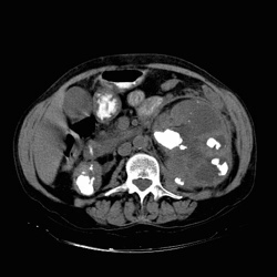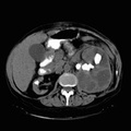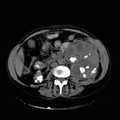
RADIOLOGY: KIDNEY: Case# 33358: ?PROBABLE XGP. 57 year old female with hematuria who is currently on hemodialysis. Patient reports that she has had numerous episodes of urinary tract infection. 1. Marked enlargement of the left kidney with dilatation of the collecting system and multi-focal calculi without presence of air. An additional focus of calcification is suspected involving the left distal ureter near the ureterovesicle junction. Mild left perinephric tissue thickening is also apparent. Prime differential considerations include malacoplakia and xanthogranulomatous pyelonephritis. A concomitant foci of renal cell carcinoma may not be totally excluded. 2. Multiple calculi within atrophic right kidney. 3. Cholelithiasis.4. Small hiatal hernia. 5. Colonic diverticula primarily sigmoid.
- Author
- Peter Anderson
- Posted on
- Thursday 1 August 2013
- Albums
- Visits
- 917


0 comments