18820/33648
)
)

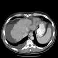
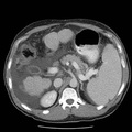
RADIOLOGY: HEPATOBILIARY: Case# 33758: HEPATOCELLULAR CARCINOMA. This is a 55-year-old male with history of cirrhosis and lesion seen on ultrasound in the left lobe of the liver. This is a follow- up with a three-phase CT study. 1. Low attenuation lesion seen during portal venous phase of contrast corresponding with the lesion seen on ultrasound. This lesion would be amenable to biopsy under ultrasound. Another lesion seen vaguely in the arterial phase in the right lobe of the liver. Hepatocellular carcinoma remains at the top of the differential for both of these lesions. 2. Varices, splenomegaly and ascites. 3. Right pleural effusion.
- Author
- Peter Anderson
- Posted on
- Thursday 1 August 2013
- Tags
- hepatobiliary, radiology
- Albums
- Visits
- 677
0 comments