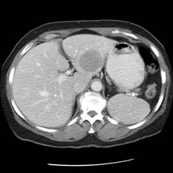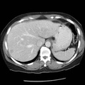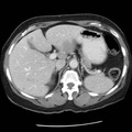
RADIOLOGY: HEPATOBILIARY: Case# 33546: CARCINOID LIVER METS. 65 year old woman. In the past she underwent left nephrectomy, but the reason for the nephrectomy is not known. 1. In addition to the well-defined 4cm low attenuation mass in the lateral segment, multiple additional lesions are seen scattered throughout the liver. These are all relatively low in attenuation compared to the liver on all phases of enhancement. These do not appear to demonstrate early arterial phase enhancement. In addition, nodes are seen in portocaval space, porta hepatis, and mesentery measuring up to 2cm in maximum dimension. The appearance is that of widespread metastatic disease. 2. Status post left nephrectomy. 3. Splenule present inferior to the spleen. The spleen, pancreas, adrenal glands and right kidney are unremarkable. 4. Myomatous uterus. 5. The lung bases are clear. Review of bone windows shows no evidence of osseous lesions.
- Author
- Peter Anderson
- Posted on
- Thursday 1 August 2013
- Tags
- carcinoid, hepatobiliary, radiology
- Albums
- Visits
- 1713


0 comments