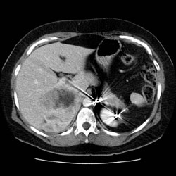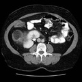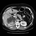
RADIOLOGY: ADRENAL: Case# 33711: MYELOLIPOMA. 46 year old female with known right adrenal mass. Evaluate tumor. 1. Large fat-containing right adrenal mass is noted which probably represent myelolipoma. The patient has a history of congenital adrenal hyperplasia and myelolipoma of the left adrenal gland. 2. Multiple low attenuation areas seen throughout the liver, the largest of which is seen in the medial segment of the left lobe of the liver measuring 2 cm represents either metastatic disease or hepatic cysts. Ultrasound could be done to look for fluid in these lesions to confirm cysts, if clinically indicated. Prior MRI & CT from 1990 were reviewed. Most of the larger lesions were seen previously. 3. Status post left adrenalectomy, cholecystecomty, and hysterectomy.
- Author
- Peter Anderson
- Posted on
- Thursday 1 August 2013
- Albums
- Visits
- 1484


0 comments