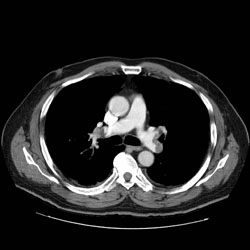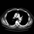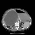
RADIOLOGY: CHEST: Case# 33706: BILATERAL PULMONARY THROMBOEMBOLI. 74 year old male with recently diagnosed diabetes mellitus who presents with left sided chest pain and abdominal pain with a 50 pound weight loss. The scan is performed to evaluate for occult malignancy. The patient has an apparent pulmonary embolus. CT Abdomen and Pelvis 1. Pulmonary arterial emboli, details which will be described on the separate CT chest report. 2. No evidence of malignancy or other significant abnormality within the abdomen or pelvis. 3. Incidental findings included small calcification in the right adrenal gland, small left renal cyst, and enlarged prostate gland. CHEST CT: 1. Bilateral pulmonary thromboemboli involving both upper and both lower lobe pulmonary arteries, greater on the right side. 2. Wedge shaped, pleural based opacities in the left upper lobe which may represent pleural effusions are probably related to the emboli. 3. Dilated right ventricle. This may also be related to the pulmonary emboli. 4. No signs of mass or lymphadenopathy.
- Author
- Peter Anderson
- Posted on
- Thursday 1 August 2013
- Albums
- Visits
- 2216


0 comments