)
)
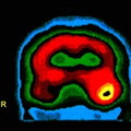
 can be calculated by dividing the mean activity in the suspicious area (mCi/ml) by the injected dose (mCi) per kilograms of body weight. Using a (SUV) of 2.5 or greater to define a malignancy, the sensitivity and specificity of 18-FDG-PET for detecting cancer in solitary pulmonary nodules greater than 1.2 cm approaches 90% with a nearly 100% specificity (1). False positives have included infectious etiologies, and sarcoid.")
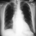
RADIOLOGY: LUNG: Case# 33670: MALIGNANT SOLITARY PULMONARY NODULE. The patient is a 67 year old woman with a solitary pulmonary nodule on a recent chest x-ray. A retrospective review of prior chest x-rays suggests that this is nodule is of recent origin. This lesion was felt to be too peripheral for reliable bronchial wash findings. Concern over potential sampling error associated with needle biopsy prompted a referral for PET imaging to rule out a malignant process. After a 4 hour fast, the patient was injected with 10 mCi of 18-FDG IV and after allowing one hour for localization, transmission and emission PET data were acquired. A hypermetabolic focus can be seen in the left upper lobe corresponding to the chest x-ray abnormality. No other abnormalities are seen. The hypermetabolic nodule suggests a malignant process without metastasis. Lesions with only slight tracer uptake can be evaluated quantitatively for significance. A significant uptake value (SUV) can be calculated by dividing the mean activity in the suspicious area (mCi/ml) by the injected dose (mCi) per kilograms of body weight. Using a (SUV) of 2.5 or greater to define a malignancy, the sensitivity and specificity of 18-FDG-PET for detecting cancer in solitary pulmonary nodules greater than 1.2 cm approaches 90% with a nearly 100% specificity (1). False positives have included infectious etiologies, and sarcoid.
- Author
- Peter Anderson
- Posted on
- Thursday 1 August 2013
- Albums
- Visits
- 1817
0 comments