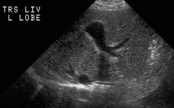
RADIOLOGY: VASCULAR: Case# 33661: PORTAL VEIN ANEURYSM. 47 year old female with right upper quadrant pain. The ultrasound images reveal dilatation of the mid to proximal portal vein with an anechoic structure that connects to the portal venous system. Pulsed Doppler demonstrates turbulent venous flow. No other abnormalities are demonstrated. Portal vein aneurysms are rare with most occurring at the junction of the superior mesenteric vein and splenic vein or more distal in the portal radicles. The etiologies include congenital or acquired secondary to portal hypertension. Most are discovered incidentally and are probably not related to the patients pain in this case.