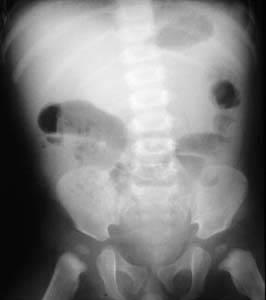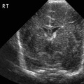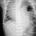
RADIOLOGY: GASTROINTESTINAL: GI: Case# 33644: INTUSSUSCEPTION. The patient is a 3-month old male with fever and abdominal pain. Plain Radiographs revealed dilated loops of bowel within the mid-abdomen. No definite distal air is identified. An ultrasound was performed to evaluate the kidneys. This revealed a loop of bowel which had a target appearance with an echogenic center and a hypoechoic rim. This was compatible with an intussusception. Barium enema was then performed to confirm the diagnosis. Barium was instilled in a retrograde fashion and a filling defect with a coiled spring appearancewas encountered in the right lower quadrant. Attempts were made to reduce this mass without success. The patient went to surgery. No pathologic leadpoint was identified. More than 90% of intussusceptions in children under 4 years of age have no demonstrable lead point. Intussusception in these children may be related to hypertrophic lymphoid tissue in the bowel wall. Intussusception can be identified and reduced under ultrasound guidance though in the United States a barium enema or air enema is more commonly performed. An enema is contraindicated when there are signs of pneumoperitoneum or peritonitis.
- Author
- Peter Anderson
- Posted on
- Thursday 1 August 2013
- Tags
- gastrointestinal, radiology
- Albums
- Visits
- 967


0 comments